Figures & data
Table 1. The correlations between MRPL23-AS1 expression and clinicopathological characteristics in 89 OS patients
Figure 1. MRPL23-AS1 is significantly increased in OS. (a,b) qRT-PCR analysis of MRPL23-AS1 in OS tissues and cell lines. (c,d) The overall and disease-free survival curves of OS patients with low or high MRPL23-AS1 expression. (e,f) qRT-PCR and FISH assays detecting the location of MRPL23-AS1 in OS cells. *p < 0.05, **p < 0.01, ***p < 0.001
Figure 2. MRPL23-AS1 promotes OS cell proliferation and invasion in vitro and in vivo. (a,b) qRT-PCR verifying the construction of stable MRPL23-AS1-overexpressing 143B cells and MRPL23-AS1-depletd U2OS cells. (c) CCK-8 assay detecting the viability of OS cells after alteration of MRPL23-AS1 level. (d,e) Transwell assay analyzing cell migration and invasion after MRPL23-AS1 knockdown or overexpression. (f) The representative image of nude mice bearing control or MRPL23-AS1-silenced U2OS cells. (g) Bioluminescence imaging showing the effect of MRPL23-AS1 knockdown on in vivo lung metastasis of U2OS cells. *p < 0.05, **p < 0.01, ***p < 0.001
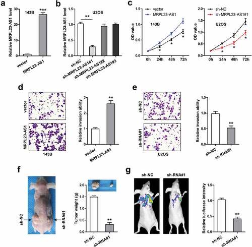
Figure 3. MRPL23-AS1 serves as a sponge of miR-30b in OS cells. (a) RNA pull-down assay using biotin-labeled MRPL23-AS1 probe, followed by qRT-PCR analysis of enrichment of miR-30b, miR-596 and miR-760. (b) The wild-type or mutant binding site between MRPL23-AS1 and miR-30b. (c) Luciferase reporter gene assay detecting the luciferase activity of wild-type or mutant MRPL23-AS1 vector after miR-30b overexpression. (d) qRT-PCR analysis of miR-30b expression after overexpression or silencing of MRPL23-AS1. (e) qRT-PCR analysis of miR-30b expression in control or MRPL23-AS1-silenced xenograft tumor tissues. (f) qRT-PCR analysis of miR-30b expression in OS and matched normal tissues. (g) The correlation between MRPL23-AS1 and miR-30b in OS tissues. (h) FISH assay showing the co-location between MRPL23-AS1 and miR-30b in OS cells. **p < 0.01, ***p < 0.001
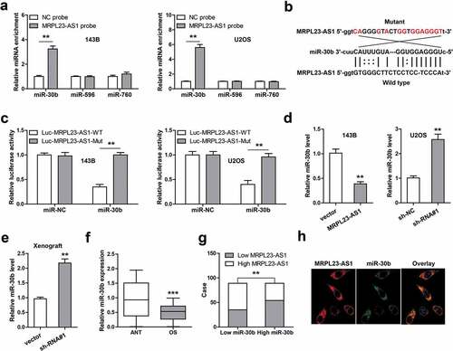
Figure 4. MRPL23-AS1 regulates the miR-30b/MYH9/Wnt/β-catenin axis in OS cells. (a) Luciferase reporter gene assay detecting the luciferase activity of wild-type or mutant MYH9 3`-UTR vector after miR-30b overexpression. (b) qRT-PCR analysis of MYH9 and β-catenin mRNA levels in MRPL23-AS1-overexpressing 143B cells after miR-30b overexpression. (c) Luciferase reporter gene assay detecting the luciferase activity of TOP/FOP-Flash reporter in MRPL23-AS1-overexpressing 143B cells after miR-30b overexpression. (d) Western blotting analyzing protein levels of MYH9, β-catenin, c-Myc and Cyclin D1, GAPDH was used as the loading control. (e) qRT-PCR analysis of MYH9 expression in OS and matched normal tissues. (f) The correlation between MRPL23-AS1 and MYH9 in OS tissues. (g,h) The relative proliferation and invasion of MRPL23-AS1-overexpressing 143B cells after transfection with miR-30b mimics, MYH9 siRNA or treatment with XAV-939. **p < 0.01, **p < 0.001. (i) The diagrammatic sketch showing MRPL23-AS1 promoting OS progression via sponging miR-30b and elevating MYH9, ultimately activating oncogenic Wnt/β-catenin pathway. **p < 0.01, ***p < 0.001