
Figures & data
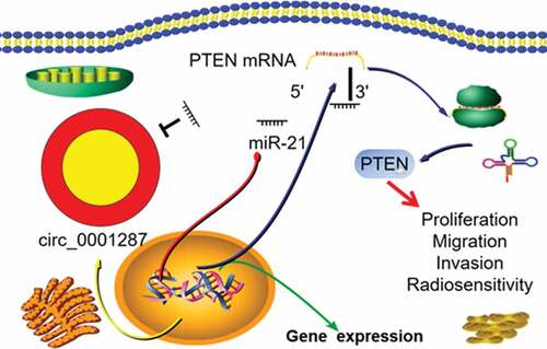
Table 1. The correlation between hsa_circ_0001287 and clinicopathological features of NSCLC patients
Figure 1. The expression of circ_0001287 in NSCLC
(a) Circ_0001287 was derived from TCAIM gene exon 6–10, with a spliced mature sequence length of 678 bp. (b) qRT-PCR was used to detect the expression level of circ_0001287 in 87 pairs of NSCLC tissues and paracancerous tissues. (c) The expression level of circ_0001287 in NSCLC cell lines (H1299, SPC-A1, A549, H2170, and H157 cells) and normal cell line (16HBE cells) was detected by qRT-PCR. The experiments were repeated three times. * P < 0.05, ** P < 0.01, and *** P < 0.001.
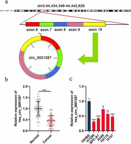
Figure 2. Circ_0001287 inhibited the multiplication, migration, invasion, and radioresistance of NSCLCs
(a) The empty plasmid or pcDNA-circ_0001287 plasmid was transfected into H1299 cells, respectively; si-NC or si-circ_0001287 was transfected into A549 cells. Transfection efficiency was verified by qRT-PCR. (b-d) The multiplication of NSCLCs was detected by CCK-8 method and BrdU experiment. (e) Transwell assay was used to detect the migration and invasion of NSCLC cells. (f) Under different doses of radiation, the survival of NSCLC cells was tested by colony formation experiments. The experiments were repeated three times. * P < 0.05, ** P < 0.01, and *** P < 0.001.
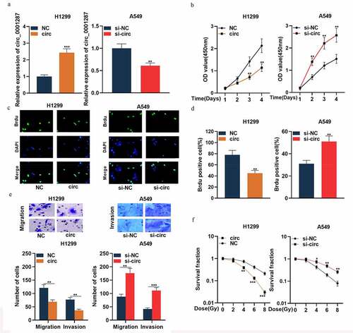
Figure 3. Circ_0001287 adsorbed miR-21
(a) Bioinformatics was used to predict the potential binding site between circ_0001287 and miR-21. (b) Wild type (WT) circ_0001287 or mutant type (MUT) circ_0001287 luciferase reporter vector and miR-con or miR-21 mimics were co-transfected into 293 T cells to measure the luciferase activity of the luciferase reporter vector. (c) RIP experiments confirmed that circ_0001287 and miR-21 were enriched in Ago2-containing microribonucleoproteins. (d) Pearson’s analysis showed that circ_0001287 expression was negatively correlated with miR-21 expression in NSCLC tissues. The experiments were repeated three times. ** P < 0.01 and *** P < 0.001.
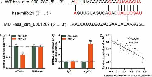
Figure 4. MiR-21 played an oncogenic role in NSCLCs
(a) The expression of miR-21 in 87 pairs of NSCLC tissues and paracancerous tissues was detected by qRT-PCR. (b) The expression of miR-21 in NSCLC cell lines (H1299, SPC-A1, A549, H2170, and H157) and normal cell line 16HBE was detected by qRT-PCR. (c) The expression of miR-21 in A549 cells transfected with miR-21 mimics and H1299 cells transfected with miR-21 inhibitors was detected by qRT-PCR. (d–e) CCK-8 method and BrdU experiment were used to detect the multiplication of NSCLC cells. (f) Transwell assay was used to monitor cell migration and invasion of NSCLC cells. (e) Under different doses of radiation, the survival of NSCLC cells was tested by colony formation experiments. The experiments were repeated three times. * P < 0.05, ** P < 0.01, and *** P < 0.001.
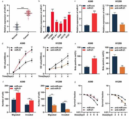
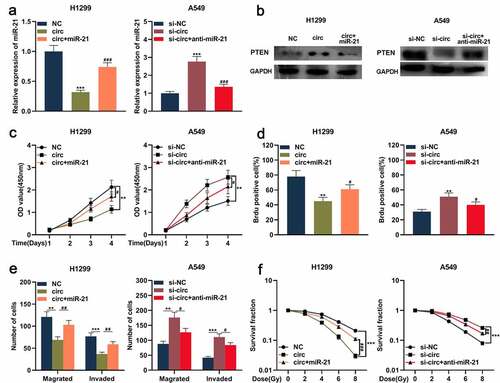
Figure 5. Circ_0001287/miR-21/PTEN axis was involved in regulating the malignant phenotype of NSCLCs
(a) miR-21 mimics and miR-21 inhibitors were transfected into the circ_0001287-H1299 cell model and the si-circ_0001287- A549 cell model, respectively. MiR-21 expression in cells was detected using qRT-PCR. (b) Western blot was used to detect the expression of PTEN protein in NSCLC cells. (c–d) Cell multiplication was detected by CCK-8 method and BrdU experiment. (e) Transwell assay was employed to detect cell migration and invasion of NSCLC cells. (f) Under different doses of radiation, the survival of NSCLC cells was tested using colony formation experiments. The experiments were repeated three times. # P < 0.05, ** ## P < 0.01, and *** ### P < 0.001.
Supplemental material
Supplemental Material
Download ()Data Availability Statement
The data used to support the findings of this study are available from the corresponding author upon request.