1,701
Views
6
CrossRef citations to date
0
Altmetric
Research Paper

Crosstalk of low density lipoprotein and liposome as a paradigm for targeting of 5-fluorouracil into hepatic cells: cytotoxicity and liver deposition

, , , , , , & show all
Pages 914-926 | Received 07 Jan 2021, Accepted 19 Feb 2021, Published online: 08 Mar 2021

Figures & data

Table 1. Particle size, zeta potential, and entrapment efficiency(EE) of the prepared liposomes

Figure 1. TEM images of plain liposomes (a), 5-FU loaded liposomes (b) 5-FUC loaded liposomes (c). (Bar = 200 nm)

Figure 1. TEM images of plain liposomes (a), 5-FU loaded liposomes (b) 5-FUC loaded liposomes (c). (Bar = 200 nm)

Figure 2. Release profiles of 5-FU and 5-FUC from liposomes. Data were expressed as mean ± SD, n = 3 in each group
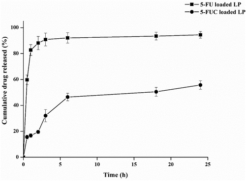
Figure 2. Release profiles of 5-FU and 5-FUC from liposomes. Data were expressed as mean ± SD, n = 3 in each group

Figure 3. SDS polyacrylamide gel electrophoresis of the LDL. Lane M, show band corresponding to standard proteins with varying molecular weights (Da). Lane 6 and 7 were samples from separated LDL
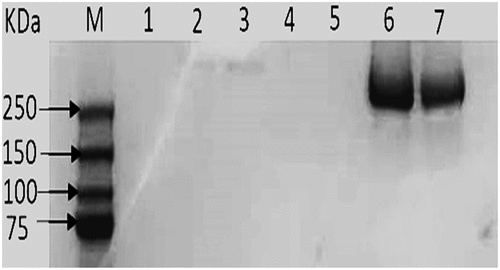
Figure 3. SDS polyacrylamide gel electrophoresis of the LDL. Lane M, show band corresponding to standard proteins with varying molecular weights (Da). Lane 6 and 7 were samples from separated LDL

Table 2. Particle size, zeta potential, and entrapment efficiency(EE) of plain LDL as well as drugs loaded LDL

Figure 4. TEM images of plain LDL (a), 5-FU loaded LDL (b) 5-FUC loaded LDL (c). (Bar = 50 nm)

Figure 4. TEM images of plain LDL (a), 5-FU loaded LDL (b) 5-FUC loaded LDL (c). (Bar = 50 nm)

Figure 5. Transfer percentage of 5-FU and 5- FUC into LDL using the liposome loading method. (a) after 2 h incubation time, (b) after 4 h incubation time
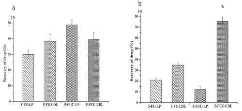
Data were expressed as mean ± SD, n = 3 in each group*, significant increase at p-value <0.05
Figure 5. Transfer percentage of 5-FU and 5- FUC into LDL using the liposome loading method. (a) after 2 h incubation time, (b) after 4 h incubation time

Figure 6. Effect of the 5-FUC solution, 5-FUC loaded liposomes, and 5-FUC loaded LDL on HepG2 line cell viability after, 24 h (a), 48 (b) 72 h (c) incubation time
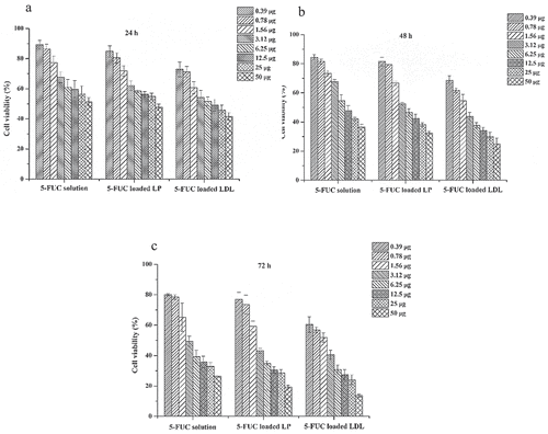
Data were expressed as mean ± SD, n = 3 in each group
Figure 6. Effect of the 5-FUC solution, 5-FUC loaded liposomes, and 5-FUC loaded LDL on HepG2 line cell viability after, 24 h (a), 48 (b) 72 h (c) incubation time

Table 3. Deposition of drugs from 5-FU and 5-FUC liposomes into LDL, and liver tissues after 2 h and 4 h of drug intraperitoneal injection