Figures & data
Figure 1. YHR inhibited the area of cardiac infarction. Heart cross section (left) and infarct area (right). Each group consists of three continuous cross sections of rat heart. The ratio of the infarct area to a single section X 100% was used as the infarct rate. The infarction rate is expressed as the mean ± SD of each group. **P <0.01, ***P < 0.001 vs. Model group
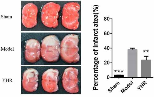
Figure 2. YHR suppressed cell apoptosis of cardiac tissue in HF model
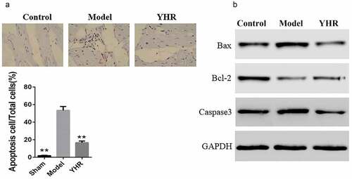
Figure 3. YHR rescued the loss of cell viability induced by OGD/R
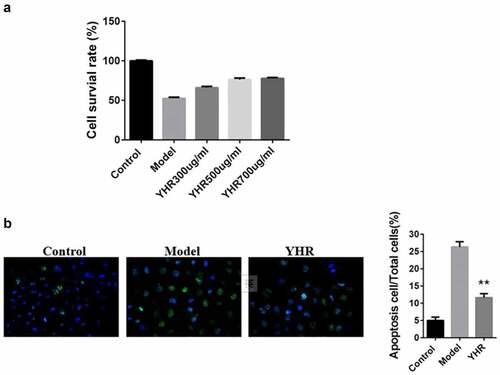
Figure 4. Detect the changes of YHR on the level of mitochondrial membrane potential (ΔΨm) of H9C2 cells induced by OGD/R
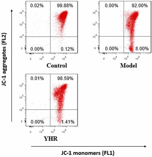
Figure 5. YHR attenuates the production of ROS in cardiomyocytes induced by OGD/R
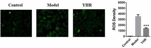
Figure 6. YHR suppressed the activation of Keap1/Nrf2/HIF-1α pathway