Figures & data
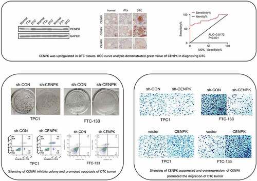
Figure 1. CENPK is upregulated in DTC tissues
Notes: (A) Expression of CENPK was significantly upregulated in PTC tissues than that in 12 normal tissue (P < 0.001). (B) Expression of CENPK was significantly upregulated in 27 FTC tissue than that in 25 FTA tissue (P < 0.001. (C) Relative mRNA level of CENPK was significantly upregulated in DTC tissues by qRT-PCR. (D) Protein level of CENPK in DTC tissues was significantly higher than that in normal thyroid tissues as detected by western blotting. (E) Immunohistochemistry staining showed that CENPK was negative in FTA and normal tissues but was highly expressed in DTC tissue. (F) ROC curve analysis for the predictive power of CENPK using qRT-PCR expression dataset. *P < 0.05, **P < 0.01, ***P < 0.001.
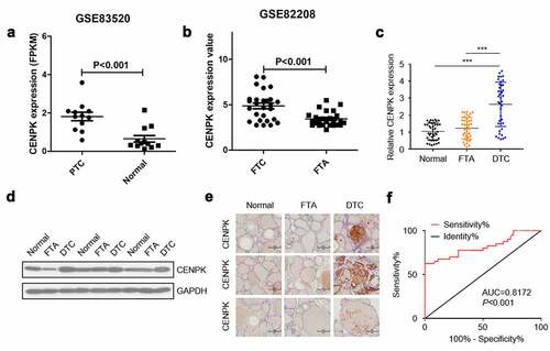
Figure 2. Silencing of CENPK inhibits DTC cell proliferation and colony
Notes: (A) Relative mRNA level of CENPK was significantly suppressed in TPC1 cells when transfected with sh-CENPK-encoding lentivirus. (B) The protein expression of CENPK was significantly suppressed in both TPC1 cells and FTC-133 cells when transfected with sh-CENPK-encoding lentivirus as shown by Western blotting. (C) The downregulation of CENPK evidently suppressed cell viability as shown by CCK-8 assay. (D) Silencing of CENPK reduced the colony number of both TPC1 cells and FTC-133 as shown by colony formation assays. (E) Knockdown of CENPK promoted apoptosis of TPC1 cells as shown by Annexin V Apoptosis Detection Kit. *P < 0.05, **P < 0.01, ***P < 0.001.
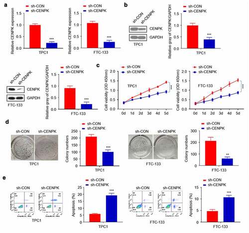
Figure 3. Overexpression of CENPK promotes DTC cell proliferation and colony
Notes: (A) Expression of CENPK was significantly elevated in both TPC1 cells and FTC-133 cells transfected with CENPK plasmid. (B) The mRNA and protein expression of CENPK was significantly elevated in both TPC1 cells and FTC-133 cells transfected with CENPK plasmid. (C) The upregulation of CENPK evidently promoted cell viability as shown by CCK-8 assay. (D) Overexpression of CENPK promoted the colony number verified by both TPC1 cells and FTC-133 cells as shown by colony formation assays. *P < 0.05, **P < 0.01, ***P < 0.001.
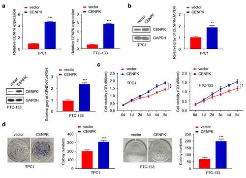
Figure 4. Influence of knockdown and overexpression of CENPK on DTC tumor migration, and invasion in vitro
Notes:(A) Silencing of CENPK suppressed the migration of both TPC1 cells and FTC-133 cells as detected by wound healing assay. (B) Silencing of CENPK suppressed the invasion of both TPC1 cells and FTC-133 cells as detected by transwell assays. (C) Overexpression of CENPK promoted migration of both TPC1 cells and FTC-133 cells. (D) Over expression of CENPK promoted invasion of TPC1 cells. *P < 0.05, **P < 0.01, ***P < 0.001.
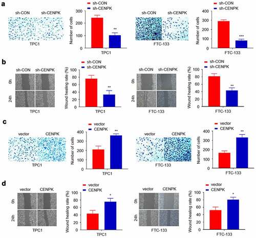
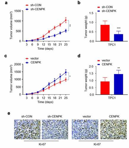
Figure 5. Influence of knockdown and overexpression of CENPK suppresses DTC tumor growth in vivo
Notes: (A) Silencing of CENPK significantly lowered the average tumor volume. (B) Silencing of CENPK significantly lowered the average tumor weight. (C) Overexpression of CENPK significantly elevated the average tumor volume. (D) Overexpression of CENPK significantly elevated the average tumor weight. (E) Silencing of CENP suppressed Ki-67 expression and Overexpression of CENPK promoted Ki-67 expression. *P < 0.05, **P < 0.01, ***P < 0.001.