
Figures & data
Figure 1. PM2.5 induces increased expression of REDD1 in BEAS-2B
(A) The viability of BEAS-2B cells after PM2.5 inducement for 6, 12, and 24 h, detected by CCK-8. (B-C) REDD1 expression in BEAS-2B cells after PM2.5 inducement for 6, 12, and 24 h, detected by RT-qPCR and Western blot. *p < 0.05, **p < 0.01, ***p < 0.001.
Figure 2. REDD1 inhibition ameliorates PM2.5-induced viability damage and inflammatory response in BEAS-2B cells
(A-B) REDD1 expression in BEAS-2B cells transfected with control, si-NC, si-REDD1-1 or si-REDD1-2, detected by RT-qPCR and Western blot. (C) The viability of PM2.5-treated BEAS-2B cells before and after transfection with si-REDD1, detected by CCK-8. (D) The expressions of pro-inflammatory cytokines TNF-α, IL-1β and IL-6 in PM2.5-treated BEAS-2B cells before and after transfection with si-REDD1, detected by RT-qPCR. (E) The expressions of proteins NF-kappaB p65 and COX-2 in PM2.5-treated BEAS-2B cells before and after transfection with si-REDD1, detected by Western blot. **p < 0.01, ***p < 0.001.
Figure 3. REDD1 inhibition attenuates PM2.5-induced BEAS-2B cell apoptosis
(A) The apoptosis of PM2.5-treated BEAS-2B cells before and after transfection with si-REDD1, observed with TUNEL staining. (B) The expressions of apoptotic proteins Bax, cleaved caspase3, cleaved caspase9, cleaved PARP and Bcl2 in PM2.5-treated BEAS-2B cells before and after transfection with si-REDD1, detected by Western blot. ***p < 0.001.
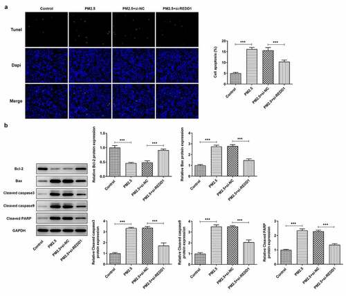
Figure 4. REDD1 interference inhibits PM2.5-induced autophagy expression in BEAS-2B cells
(A) The expressions of autophagy-related proteins in BEAS-2B cells after PM2.5 inducement for 6, 12, and 24 h, detected by Western blot. (B) The expressions of autophagy-related proteins in PM2.5-treated BEAS-2B cells before and after transfection with si-REDD1, detected by Western blot. (C) The expression of p-mTOR and mTOR in PM2.5-treated BEAS-2B cells before and after transfection with si-REDD1, detected by Western blot. (D) The autophagy level of PM2.5-treated BEAS-2B cells before and after transfection with si-REDD1, observed with GFP-LC3 staining. *p < 0.05, **p < 0.01, ***p < 0.001.
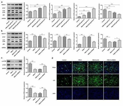
Figure 5. REDD1 interference ameliorates PM2.5-induced viability damage and inflammatory injury in BEAS-2B cells via inhibiting autophagy
(A) The viability of BEAS-2B cells treated with control, PM2.5, PM2.5+ si-REDD1 or PM2.5+ si-REDD1+ rapamycin, detected by CCK-8. (B) The expressions of pro-inflammatory cytokines TNF-a, IL-1β and IL-6 in BEAS-2B cells treated with control, PM2.5, PM2.5+ si-REDD1 or PM2.5+ si-REDD1+ rapamycin, detected by RT-qPCR. (C) The expressions of NF-kappaB p65 and COX-2 in BEAS-2B cells treated with control, PM2.5, PM2.5+ si-REDD1 or PM2.5+ si-REDD1+ rapamycin, detected by Western blot. *p < 0.05, **p < 0.01, ***p < 0.001.
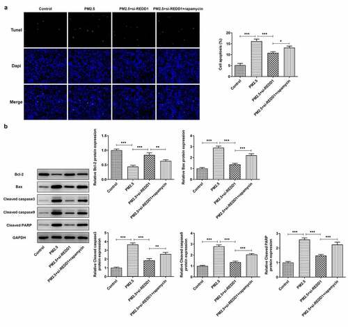
Figure 6. REDD1 interference ameliorates PM2.5-induced BEAS-2B cell apoptosis via inhibiting autophagy
(A) The apoptosis of BEAS-2B cells treated with control, PM2.5, PM2.5+ si-REDD1 or PM2.5+ si-REDD1+ rapamycin, observed with TUNEL staining. (B) The expressions of Bax, cleaved caspase3, cleaved caspase9, cleaved PARP and Bcl2 in BEAS-2B cells treated with control, PM2.5, PM2.5+ si-REDD1 or PM2.5+ si-REDD1+ rapamycin, detected by Western blot. *p < 0.05, **p < 0.01, ***p < 0.001.
Availability of data
The datasets used and analyzed are available from the corresponding author and first author on reasonable request.