Figures & data
Table 1. General clinical data of the patients in the two groups (mean ± SD)
Figure 1. Fus is up-regulated in plasma of AF patients and AngII-induced cardiac fibroblasts. (a), the expression of FUS in plasma of patients with sinus rhythm (control, n = 30) and atrial fibrillation (AF, n = 30) was detected by RT-qPCR. (b and c), the mRNA (b) and protein (c) expressions of Fus in control mouse cardiac fibroblasts and cells that stimulated with 1 μM AngII for 12 h. **P < 0.01 and ***P < 0.001
Figure 2. Fus knockdown inhibits AngII-induced proliferation, migration and collagen synthesis of cardiac fibroblasts. (a and b), the mRNA (a) and protein (b) expressions of Fus in control mouse cardiac fibroblasts and cells that transfected with indicated shRNAs. ***P < 0.001. (c–e), mouse cardiac fibroblasts that silenced with Fus or not were stimulated with 1 μM AngII for 12 h, then (c) the cell viability was measured by CCK-8 assay, ***P < 0.001 vs Control, ###P < 0.001 vs AngII + sh-NC; (d) cell migration was detected by wound healing assay; (e) the protein expression of α-SMA, collagen 1 and fibronectin was assessed by western blot. *P < 0.05 and ***P < 0.001
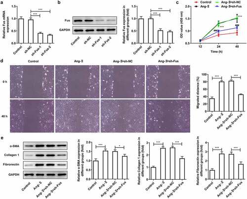
Figure 3. Fus knockdown inhibits AngII-induced TGF-β1/Smad pathway activation. Mouse cardiac fibroblasts that silenced with Fus or not were stimulated with 1 μM AngII for 12 h, then the protein expression of TGF-β1, phosphorylated (p)-Smad2/Smad2 and p-Smad3/Smad3 was assessed by western blot. **P < 0.01 and ***P < 0.001
Figure 4. Fus binds to Pax3 and regulates Pax3 expression. (a and b), the interaction between Fus and Pax3 mRNA in mouse cardiac fibroblasts was verified by RIP (a) and RNA pull down (b) assays. (c and d), mouse cardiac fibroblasts that silenced with Fus or not were stimulated with 1 μM AngII for 12 h, then the mRNA (c) and protein (d) expressions of Pax3 were measured by RT-qPCR and western blot. *P < 0.05, **P < 0.01 and ***P < 0.001
Figure 5. Pax3 overexpression blocks the effect of Fus knockdown on AngII-induced TGF-β1/Smad pathway activation. (a), the protein expression of Pax3 in mouse cardiac fibroblasts before and after Pax3 overexpression. (b), mouse cardiac fibroblasts that co-transfected with shRNA-Fus and pcDNA-Pax3 or not were stimulated with 1 μM AngII for 12 h, then the protein expression of TGF-β1, phosphorylated (p)-Smad2/Smad2 and p-Smad3/Smad3 was assessed by western blot. *P < 0.05, **P < 0.01 and ***P < 0.001
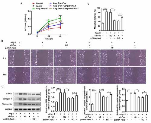
Figure 6. Pax3 overexpression blocks the effect of Fus knockdown on AngII-induced proliferation, migration and collagen synthesis of cardiac fibroblasts. Mouse cardiac fibroblasts that co-transfected with shRNA-Fus and pcDNA-Pax3 or not were stimulated with 1 μM AngII for 12 h, then (a) the cell viability was measured by CCK-8 assay, ***P < 0.001 vs Control, ###P < 0.001 vs AngII + sh-NC, ΔP<0.05 vs AngII + sh-Fus + pcDNA3.1; (b and c) cell migration was detected by wound healing assay; (d) the protein expression of α-SMA, collagen 1 and fibronectin was assessed by western blot. *P < 0.05 and ***P < 0.001