Figures & data
Table 1. The sequence of primers used in RT-qPCR
Figure 1. FTO and STAT3 expression in doxorubicin-resistant BC cells. a, b The sensitivity of doxorubicin-resistant MCF-7 and MDA-MB-231 cells and their parental cells to doxorubicin. c The IC50 value of doxorubicin against doxorubicin-resistant MCF-7 and MDA-MB-231 cells and their parental cells. d, e The mRNA level of FTO and STAT3 in doxorubicin-resistant BC cells and their parental cells. f, g The protein expression of FTO, p-STAT3 and STAT3 in doxorubicin-resistant BC cells and their parental cells. **p < 0.01. MCF-7-DoxR, doxorubicin-resistant MCF-7; MDA-MB-231-DoxR, doxorubicin-resistant MDA-MB-231; IC50, half-maximal (50%) inhibitory concentration; p-STAT3, phosopho-STAT3Tyr705
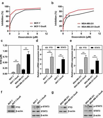
Figure 2. Effect of STAT3 on the transcriptional activity of FTO promoter and the expression of FTO in doxorubicin-resistant BC cells and their parental cells. a The binding sites of STAT3 in FTO promoter predicted by JASPAR. b CHIP assay confirmed the binding between FTO promoter and STAT3 in doxorubicin-resistant MCF-7 cells. c, d Dual luciferase reporter assay confirmed that the transcriptional activity of FTO promoter was inhibited by S3I-201 but enhanced by EGF in doxorubicin-resistant MCF-7 cells and MCF-7 cells. e-l The mRNA and protein expression of FTO was evaluated using RT-qPCR and western blotting. *p < 0.05, **p < 0.01. MCF-7-DoxR, doxorubicin-resistant MCF-7; MDA-MB-231-DoxR, doxorubicin-resistant MDA-MB-231; EGF, epidermal growth factor
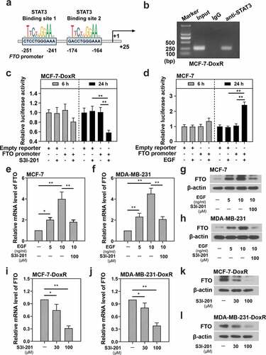
Figure 3. Effect of FTO on the activation of STAT3 signaling in doxorubicin-resistant BC cells and their parental cells. Doxorubicin-resistant BC cells and their parental cells were transfected with FTO shRNAs and FTO overexpression plasmids for 72 h, respectively. a-d The protein and mRNA expression of FTO in transfected and untransfected cells. e, f Total m6A level in transfected and untransfected cells. g-j The protein expression of p-STAT3 and STAT3 and the mRNA expression of STAT3 in transfected and untransfected cells. **p < 0.01. MCF-7-DoxR, doxorubicin-resistant MCF-7; MDA-MB-231-DoxR, doxorubicin-resistant MDA-MB-231; shNC, negative control shRNA; shFTO, FTO shRNA; NC-OE, empty vector; OE-FTO, FTO overexpression; p-STAT3, phosopho-STAT3Tyr705; m6A, N6-methyladenosine
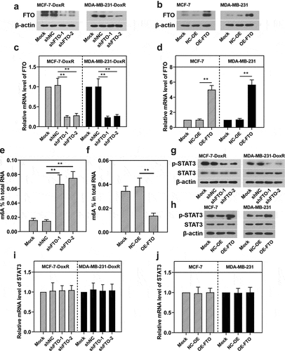
Figure 4. Role of STAT3-FTO in the doxorubicin-induced loss of BC cell viability. Doxorubicin-resistant BC or BC cells were transfected with FTO siRNAs or FTO overexpression plasmids and co-transfected with FTO siRNA and FTO overexpression plasmids for 48 h and then exposed to doxorubicin for 24 h. a-f Cell viability was measured by CCK-8 assay. *p < 0.05, **p < 0.01. MCF-7-DoxR, doxorubicin-resistant MCF-7; MDA-MB-231-DoxR, doxorubicin-resistant MDA-MB-231; siNC, negative control siRNA; siFTO, FTO siRNA; NC-OE, empty vector; OE-FTO, FTO overexpression; OD, optical density
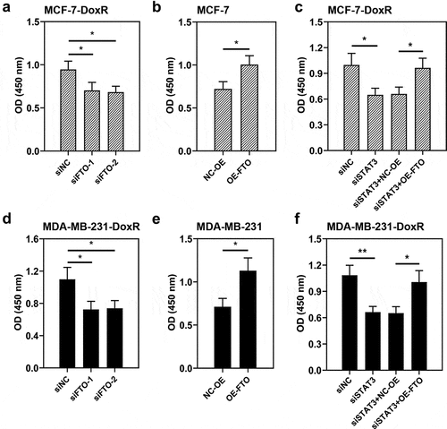
Figure 5. Role of STAT3-FTO in the doxorubicin-induced apoptosis of MCF-7 cells. Doxorubicin-resistant MCF-7 or MCF-7 cells were transfected with FTO siRNAs or FTO overexpression plasmids and co-transfected with FTO siRNA and FTO overexpression plasmids for 48 h and then exposed to doxorubicin for 24 h. a, b and d Apoptotic MCF-7 cells were detected by Annexin V/PI staining. c, e Expressions of cleaved PARP and cleaved caspase-3 in doxorubicin-resistant MCF-7 and MCF-7 cells. **p < 0.01. MCF-7-DoxR, doxorubicin-resistant MCF-7; siNC, negative control siRNA; siFTO, FTO siRNA; NC-OE, empty vector; OE-FTO, FTO overexpression; PI, propidium iodide; PARP, poly (ADP-ribose) polymerase
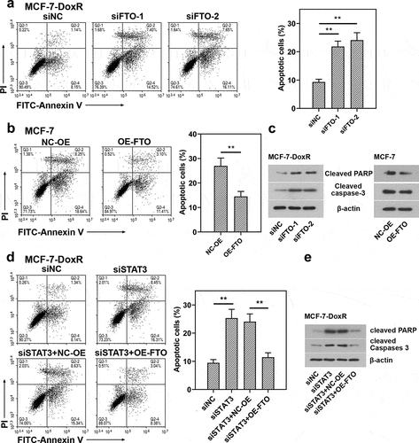
Figure 6. Role of STAT3-FTO in the doxorubicin-induced apoptosis of MDA-MB-231 cells. Doxorubicin-resistant MDA-MB-231 or MDA-MB-231 cells were transfected with FTO siRNAs or FTO overexpression plasmids and co-transfected with FTO siRNA and FTO overexpression plasmids for 48 h and then exposed to doxorubicin for 24 h. a, b and d Apoptotic MDA-MB-231 cells were detected by Annexin V/PI staining. c, e Expressions of cleaved PARP and cleaved caspase-3 in doxorubicin-resistant MDA-MB-231 and MDA-MB-231 cells. **p < 0.01. MDA-MB-231-DoxR, doxorubicin-resistant MDA-MB-231; siNC, negative control siRNA; siFTO, FTO siRNA; NC-OE, empty vector; OE-FTO, FTO overexpression; PI, propidium iodide; PARP, poly (ADP-ribose) polymerase

Figure 7. Effect of doxorubicin on the viability and apoptosis of Hs578T cells. Hs578T cells were exposed to doxorubicin for 24 h. a Cell viability was measured by CCK-8 assay. b Apoptotic Hs578T cells were detected by Annexin V/PI staining. *p < 0.05, **p < 0.01. Dox, doxorubicin; OD, optical density; PI, propidium iodide
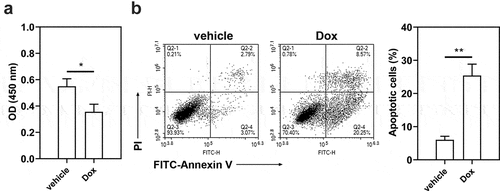
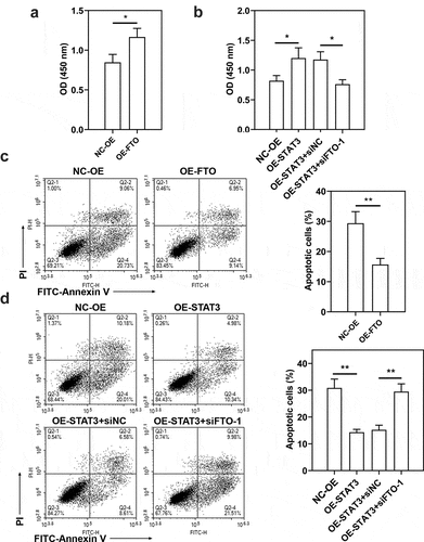
Figure 8. Role of STAT3-FTO in the doxorubicin-induced viability loss and apoptosis of Hs578T cells. Hs578T cells were transfected with FTO overexpression plasmids and co-transfected with STAT3 overexpression plasmids and FTO siRNA-1 for 48 h and then exposed to doxorubicin for 24 h. a, b Cell viability was measured by CCK-8 assay. c, d Apoptotic Hs578T cells were detected by Annexin V/PI staining. *p < 0.05, **p < 0.01. Dox, doxorubicin; OD, optical density; PI, propidium iodide; NC-OE, empty vector; OE-FTO, FTO overexpression; OE-STAT3, STAT3 overexpression; siNC, negative control siRNA; siFTO-1, FTO siRNA-1