Figures & data
Figure 1. Cell viability under different concentrations of ginsenoside Rg3
The SMMC-2271 cells were treated with 0, 1, 2, 4, 8 and 16 μg/ml ginsenoside Rg3 and the cell viability was evaluated by MTT assay after incubation.
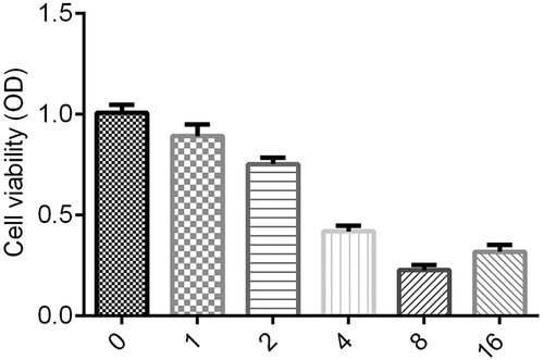
Figure 2. Reduced lncRNA-HOTAIR expression by ginsenoside Rg3
SMMC-7721 and SK-Hep-1 cells were incubated with 8 μg/ml ginsenoside Rg3 in RPMI-1640 (600 µl) containing 10% FBS at 37°C with 5% CO2. The relative expression of lncRNA-HOTAIR was evaluated by qRT-PCR. Control: non-treated group; ginsenoside Rg3: cells treated with 8 μg/ml ginsenoside Rg3. **p < 0.05 vs. control group.
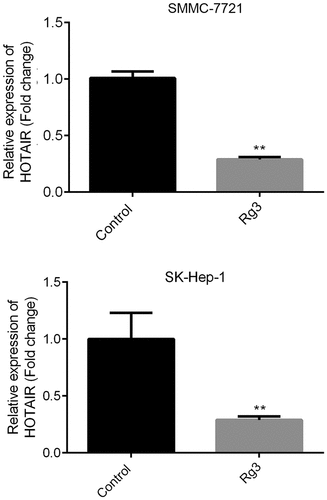
Figure 3. LncRNA-HOTAIR overexpression system
The lncRNA-HOTAIR-negative and overexpression plasmids were added to SMMC-7721 cells and incubate with RPMI-1640 (600 µl) containing 10% FBS at 37°C with 5% CO2 for 48 h. The relative expression of lncRNA-HOTAIR was determined by qRT-PCR. Control: non-treated group; pcDNA3.1: cells treated with lncRNA-HOTAIR-negative control plasmids; pcDNA3.1-HOTAIR: cells treated with lncRNA-HOTAIR overexpression plasmids. **p < 0.05 vs. control group.
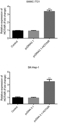
Figure 4. Inhibited cell proliferation rate by ginsenoside Rg3
The cell viability was determined by MTT assay after 0, 12, 24 and 48 h incubation with RPMI-1640 (600 µl) containing 10% FBS at 37°C with 5% CO2. Control: non-treated group; Ginsenoside Rg3: cells treated with 8 μg/ml ginsenoside Rg3. Rg3+ pcDNA3.1: cells treated with 8 μg/ml ginsenoside Rg3 and lncRNA-HOTAIR-negative control plasmids; Rg3+ lncRNA-HOTAIR-negative control plasmids: 8 μg/ml ginsenoside Rg3 and lncRNA-HOTAIR overexpression plasmids.
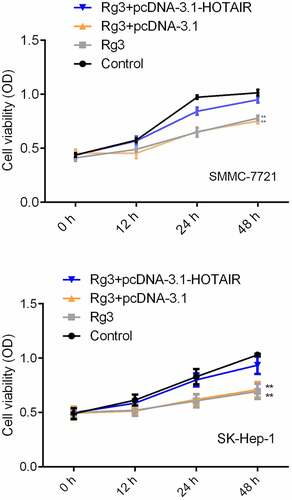
Figure 5. Inhibited cell migration by ginsenoside Rg3
(a)–(b): The cell migration ability was determined by scratch assay after 48 incubation with RPMI-1640 (600 µl) containing 10% FBS at 37°C with 5% CO2. Control: non-treated group; Ginsenoside Rg3: cells treated with 8 μg/ml ginsenoside Rg3. Rg3+ pcDNA3.1: cells treated with 8 μg/ml ginsenoside Rg3 and lncRNA-HOTAIR-negative control plasmids; Rg3+ lncRNA-HOTAIR-negative control plasmids: 8 μg/ml ginsenoside Rg3 and lncRNA-HOTAIR overexpression plasmids.
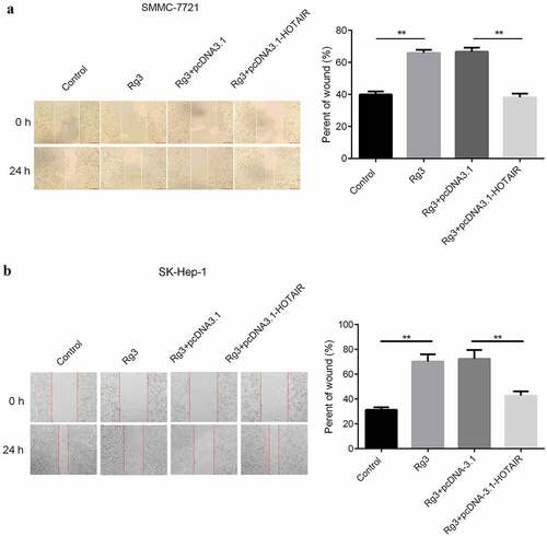
Figure 6. Inhibited cell invasion ability by ginsenoside Rg3
(a)–(b): The invasion ability was determined by a transwell assay after 48 incubation with RPMI-1640 (600 µl) containing 10% FBS at 37°C with 5% CO2. Control: non-treated group; Ginsenoside Rg3: cells treated with 8 μg/ml ginsenoside Rg3. Rg3+ pcDNA3.1: cells treated with 8 μg/ml ginsenoside Rg3 and lncRNA-HOTAIR-negative control plasmids; Rg3+ lncRNA-HOTAIR-negative control plasmids: 8 μg/ml ginsenoside Rg3 and lncRNA-HOTAIR overexpression plasmids.
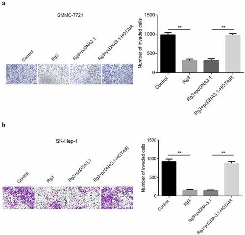
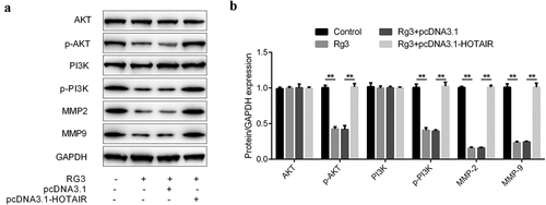
Figure 7. Inhibited expression of MMP2, MMP9, p-AKT, and p-PI3K by ginsenoside Rg3
(a): The protein level of MMP2, MMP9, PI3K/AKT, p-AKT and p-PI3K was determined by Western blot. Expression of proteins by Western blot. (b): Quantitative analysis of (a). Control: non-treated group; Ginsenoside Rg3: cells treated with 8 μg/ml ginsenoside Rg3. Rg3+ pcDNA3.1: cells treated with 8 μg/ml ginsenoside Rg3 and lncRNA-HOTAIR-negative control plasmids; Rg3+ lncRNA-HOTAIR-negative control plasmids: 8 μg/ml ginsenoside Rg3 and lncRNA-HOTAIR overexpression plasmids. **p < 0.05 vs. control group. ## p < 0.05 vs. Rg3+ lncRNA-HOTAIR-negative control group.