
Figures & data
Figure 1. The lncRNA CRNDE expression was up-regulated in PC. RT-PCR analysis was performed to measure the expression of lncRNA CRNDE in PC tissues (a) and cells (b).*p < 0.05, **p < 0.01, ***p < 0.001. Each cell experiment was repeated for 3 times
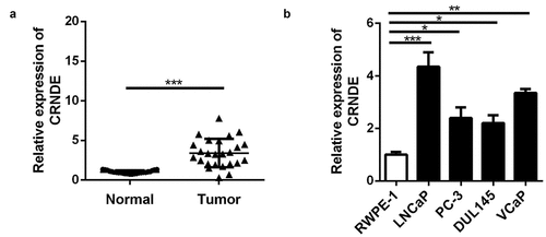
Figure 2. miR-146a-5p is a target of lncRNA CRNDE. (a) The predicted lncRNA CRNDE binding site in the miR-146a-5p 3ʹ-UTR . (b) Relative luciferase activity of cells after co-transfection with wild type (WT) or mutant (Mut) lncRNA CRNDE 3′-UTR reporter genes and miR-146a-5p mimics. (d) miR-146a-5p was up-regulated in PC tissues. (D) miR-146a-5p was up-regulated in all 4 PC cells in comparison with the RWPE-1 cells. **p < 0.01, ***p < 0.001. Each cell experiment was repeated for 3 times
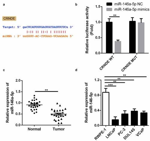
Figure 3. SiRNA and inhibitor down-regulated the expressions of CRNDE and miR-146a-5p. (a) The expression of CRNDE was detected by qRT-PCR after siRNA 1# and siRNA 2# transfection. (b) The expression of miR-146a-5p was measured by qRT-PCR after inhibitor transfection. ***p < 0.001. Each cell experiment was repeated for 3 times
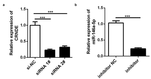
Figure 4. Knockdown of miR-146a-5p reversed the effects of si-CRNDE on the proliferation of PC. CCK-8(a) and colony formation (b) assays were used to detect the proliferation of PC3 cell lines. *p < 0.05, ***p < 0.001. Each experiment was repeated for 3 times
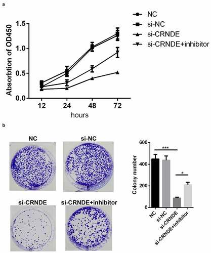
Figure 5. Knockdown of miR-146a-5p reversed the effect of si-CRNDE on the apoptosis rates of PC. (a-b) Flow cytometry assay was performed to determine the apoptosis rates of PC. **p < 0.01, ***p < 0.001. Each experiment was repeated for 3 times
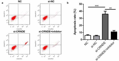
Figure 6. Knockdown of miR-146a-5p reversed the effect of si-CRNDE on the migration and invasion of PC. The number of migratory and invasive cells was determined by transwell chamber assays. Magnification ×200. **p < 0.01. Each experiment was repeated for 3 times
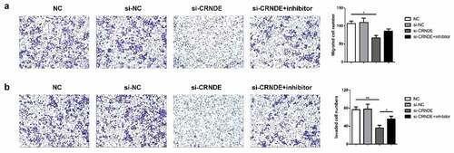
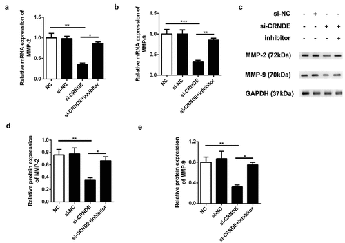
Figure 7. Knockdown of miR-146a-5p reversed the effect of si-CRNDE on the MMP-2 and MMP-9 expressions of PC. The qRT-PCR (a-b) and western blot (c-e) assays to detect the expressionS of MMP-2 and MMP-9 at mRNAs and proteins levels. **p < 0.01. Each experiment was repeated for 3 times
Availability of data and materials
The datasets used and/or analyzed during the current study are available from the corresponding author on reasonable request.