Figures & data
Figure 1. The pathological state of MAS rats was significantly alleviated by GA. A. The W/D ratio of lung tissues was calculated. B. The concentration of IL-6, IL-1β, and TNF-α was detected by ELISA assay. C. HE staining was used to detect the pathological state of lung tissues (**p < 0.01 vs. Control, #p < 0.05 vs. Model, ##p < 0.01 vs. Model)
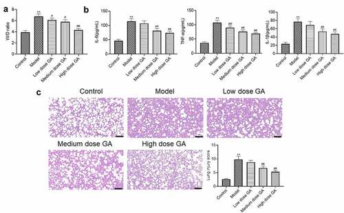
Figure 2. The apoptotic state of lung tissues from MAS rats was significantly mitigated by GA. A. The apoptotic index of lung tissues was evaluated by TUNEL assay. B. The expression of Bcl-2, Bax, and cleaved-Caspase-3 was determined by Western blotting assay (**p < 0.01 vs. Control, #p < 0.05 vs. Model, ##p < 0.01 vs. Model)
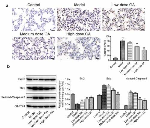
Figure 3. The oxidative stress in WAS rats was dramatically suppressed by GA. A. The concentration of MDA, SOD, and GSH was determined by ELISA assay. B. The expression of Nrf2, Keap1, and HO-1 was determined by Western blotting assay (**p < 0.01 vs. Control, #p < 0.05 vs. Model, ##p < 0.01 vs. Model)
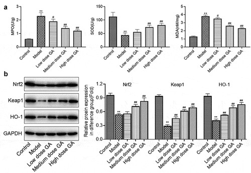
Figure 4. The protective effect of GA on the pathology of WAS tissues was abolished by ML385. A. The W/D ratio of lung tissues was calculated. B. The concentration of IL-6, IL-1β, and TNF-α was detected by ELISA assay. C. HE staining was used to detect the pathological state of lung tissues (**p < 0.01 vs. Control, ##p < 0.01 vs. Model, &p < 0.05 vs. GA, &&p < 0.01 vs. GA)
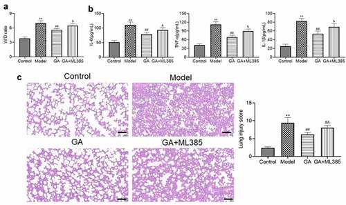
Figure 5. The protective property of GA on the apoptotic state of WAS tissues was abolished by ML385. A. The apoptotic index of lung tissues was evaluated by TUNEL assay. B. The expression level of Bcl-2, Bax, and cleaved-Caspase-3 was determined by Western blotting assay (**p < 0.01 vs. Control, ##p < 0.01 vs. Model, &p < 0.05 vs. GA, &&p < 0.01 vs. GA)
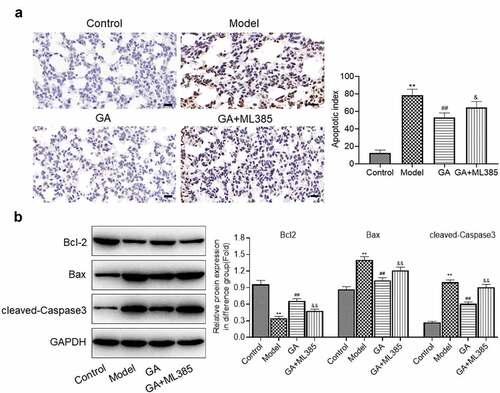
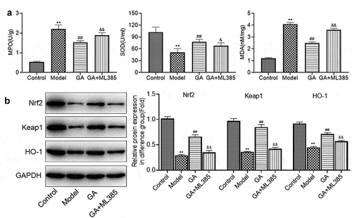
Figure 6. The protective effect of GA on oxidative stress of WAS tissues was abolished by ML385. A. The concentration of MDA, SOD, and GSH was determined by ELISA assay. B. The expression of Nrf2, Keap1, and HO-1 was determined by Western blotting assay (**p < 0.01 vs. Control, ##p < 0.01 vs. Model, &p < 0.05 vs. GA, &&p < 0.01 vs. GA)