Figures & data
Figure 1. Cytotoxicity of Telmisartan in bronchial BEAS-2B epithelial cells. (a-b). Cells were incubated with Telmisartan at concentrations of 1, 2, 10, 20, 100, and 200 μM for 24 hours. Cell viability normalized to vehicle group while LDH release normalized to total LDH in each group (%); (c-d). Cells were challenged with LPS (30 μg/ml) with or without Telmisartan (10, and 20 μM) for 24 hours. Cell viability normalized to vehicle group while LDH release normalized to total LDH in each group (%) (*, **, ****, P < 0.05, 0.01, 0.0001 vs. vehicle group; ##, ###, P < 0.01, 0.001 vs. LPS group)
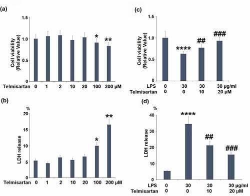
Figure 2. Telmisartan improved LPS-induced mitochondrial dysfunction in bronchial BEAS-2B epithelial cells. Cells were challenged with LPS (30 μg/ml) with or without Telmisartan (10, and 20 μM) for 24 hours. (a). Levels of mitochondrial membrane potential (ΔΨm) normalized to vehicle group; (b). level of cellular ATP normalized to vehicle group (****, P < 0.0001 vs. vehicle group; ##, ###, P < 0.01, 0.001 vs. LPS group)
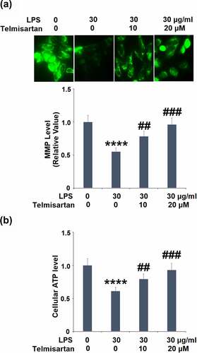
Figure 3. Telmisartan suppressed LPS-induced expression and production of proinflammatory cytokines. Cells were challenged with LPS (30 μg/ml) with or without Telmisartan (10, and 20 μM) for 24 hours. (a-c). mRNA of TNF-α, IL-6, and TGF-β normalized to control group; (d-f). Production of TNF-α, IL-6, and TGF-β (****, P < 0.0001 vs. vehicle group; ##, ###, P < 0.01, 0.001 vs. LPS group)
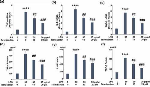
Figure 4. Telmisartan suppressed LPS-induced production of MUC5AC. Cells were challenged with LPS (30 μg/ml) with or without Telmisartan (10, and 20 μM) for 24 hours. (a). mRNA of MUC5AC normalized to vehicle group; (b). Production of MUC5AC (****, P < 0.0001 vs. vehicle group; ##, ###, P < 0.01, 0.001 vs. LPS group)
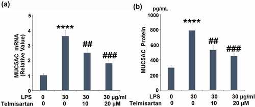
Figure 5. Telmisartan alleviated LPS-induced decrease in the expression of SOCS1. Cells were challenged with LPS (30 μg/ml) with or without Telmisartan (10, and 20 μM) for 24 hours. (a). mRNA of SOCS1 normalized to vehicle group; (b). Protein of SOCS1 normalized to vehicle group (****, P < 0.0001 vs. vehicle group; ##, ###, P < 0.01, 0.001 vs. LPS group)
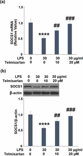
Figure 6. Telmisartan inhibited LPS-induced activation of NF-κB. Cells were transfected with NF-κB dependent luciferase expression plasmid, followed by stimulation with LPS (30 μg/ml) with or without Telmisartan (10, and 20 μM) for 24 hours. Luciferase activity was measured (****, P < 0.0001 vs. vehicle group; ##, ###, P < 0.01, 0.001 vs. LPS group)
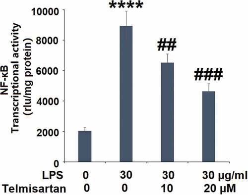
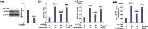
Figure 7. Silencing of SOCS1 abolished the protective effects of Telmisartan against LPS- induced production of MUC5AC. Cells were transfected with SOCS1 siRNA, followed by challenging with LPS (30 μg/ml) with or without Telmisartan (10, and 20 μM) for 24 hours. (a). Western blot analysis revealed successful knockdown of SOCS1; (b). mRNA of MUC5AC; (c). Production of MUC5AC; (d) Transcriptional activity of NF-kB measured by a luciferase assay (****, P < 0.0001 vs. vehicle group; ###, P < 0.001 vs. LPS group; $$$, P < 0.001 vs. LPS+ Telmisartan group)