Figures & data
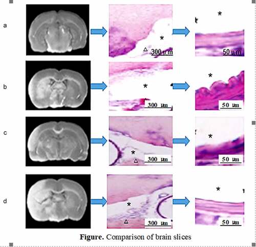
Table 1. Zeal-Longa five-point scale scoring method
Figure 1. Results of evaluation of neurologic impairment
(Note: *represented a significant difference from group A (control group), P < 0.05; # represented a significant difference from group A and group C at the same time, P < 0.05.)
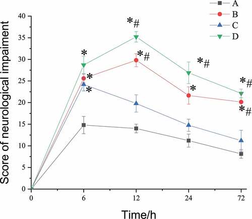
Figure 2. Comparison of number of micro-vasoconstrictions after operation
(* meant that there was statistically considerable difference, P < 0.05.)
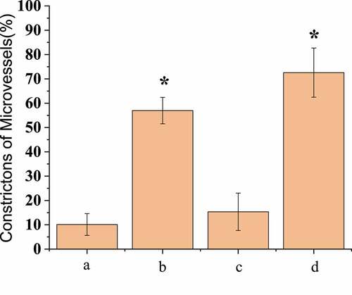
Figure 3. Comparison of the degree of micro-vasoconstriction after operation
(* meant that there was statistically considerable difference, P < 0.05.)
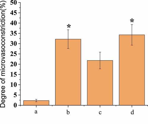
Figure 4. Fluorescence imaging in vivo
(Note: the yellow circle in the figure indicated the artery lumen.)
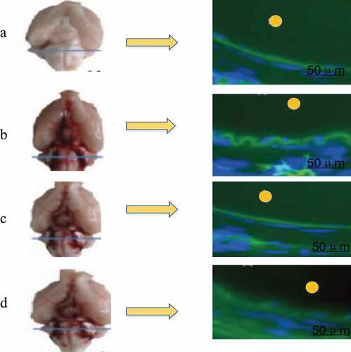
Figure 5. BBB permeability test results
(* meant that there was statistically considerable difference, P < 0.05.)
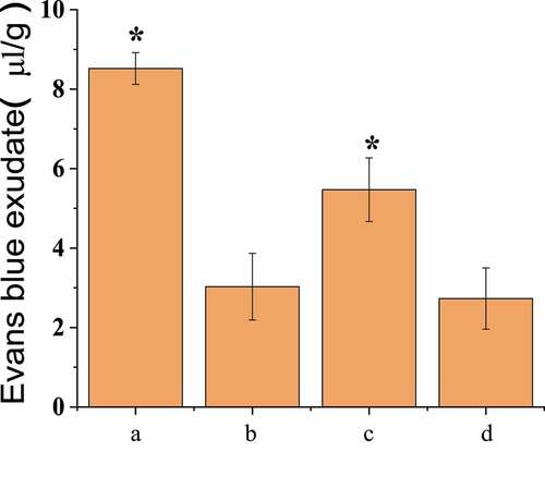
Figure 6. Comparison of brain slices
(Note: * in the figure represented the arterial lumen; the triangles represented blood clots in the subarachnoid space; the middle column was the general image, and the last column was the partial enlarged image.)
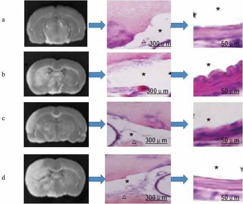
Figure 7. Comparison of the expression results of α-SMA in pericytes of each group
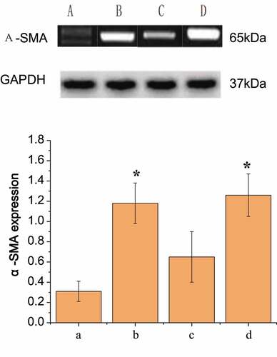
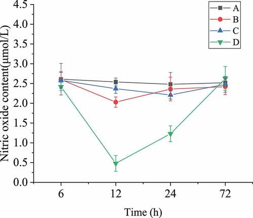
Figure 8. Nitric oxide content in blood of rats after SAH operation