Figures & data
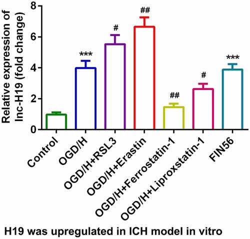
Figure 1. H19 was highly expressed in the OGD/H ICH model cells
(a) Expression of H19 in BMVECs. (b) Levels of GSH in BMVECs. (c) Levels of iron in BMVECs. (d) Levels of LPO in BMVECs. (e) Levels of MDA in BMVECs.**P < 0.01, ***P < 0.001, compared with the control group. #P < 0.05, ##P < 0.01, compared with the OGD/H group.
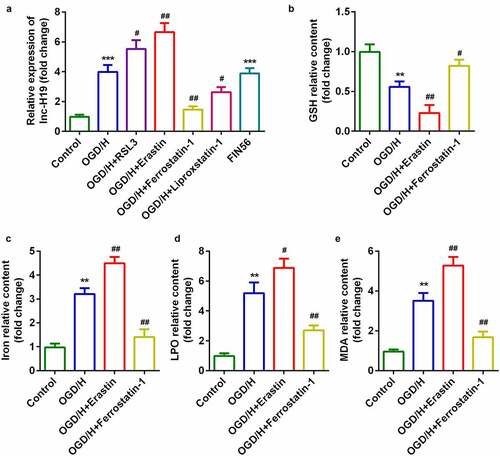
Figure 2. Knockdown of H19 promoted cell viability and inhibited ferroptosis
(a) H19 expression levels were detected using RT-qPCR after H19 knockdown. (b) Cell viability was measured using CCK8 assays after transfection with si-H19. (c) PI staining was used to detect cell death. (d) TUNEL staining was used to detect cell death. (e–h) Levels of iron, GSH, MDA, and LPO. (i–k) Expression of SLC7A11, GPX4, and TFR1 mRNAs were measured using RT-qPCT. (l) Expression of SLC7A11, GPX4, and TFR1 proteins detected by western blotting.*P < 0.05, **P < 0.01, compared with si-NC group or control group. #P < 0.05, ##P < 0.01, compared with the OGD/H group. &P < 0.05, &&P < 0.01, compared with the OGD/H + Ferrostatin + si-NC group.
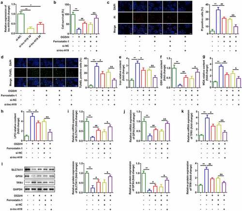
Figure 3. H19 sponged miR-106b-5p in BMVECs
(a) Bioinformatic analysis predicted the binding sites between miR-106b-5p and H19. (b) Dual-luciferase reporter assays confirmed that miR-106b-5p was a target of H19 in BMVECs. (c) Interactions between miR-106b-5p and H19 were determined using RNA pull-down assays. *P < 0.05 (d) The expression of miR-106b-5p was determined by RT-qPCR. (E) miR-106b-5p expression levels in BMVECs treated with or without OGD/H. *P < 0.05, **P < 0.01, ##P < 0.01.
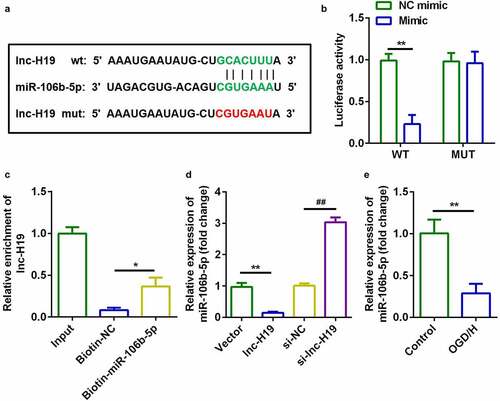
Figure 4. Down-regulation of miR-106b-5p reversed the effects of H19 knockdown on cell viability and ferroptosis of BMVECs
(a) miR-106b-5p expression was detected using RT-qPCR after H19`knockdown. (b) Cell viability was detected using CCK8 assays after transfection with si-H19. (c) PI staining was used to detect cell death. (d) TUNEL staining was used to detect cell death. (E-H) Levels of iron, GSH, MDA, and LPO. (i–k) Levels of SLC7A11, GPX4, and TFR1 mRNAs were measured using RT-qPCT. (l) Expression of SLC7A11, GPX4, and TFR1 proteins detected by western blotting. **P < 0.01, compared with NC inhibitor group or control group. #P < 0.05, ##P < 0.01, compared with OGD/H + si-lnc-H19 + NC inhibitor group.
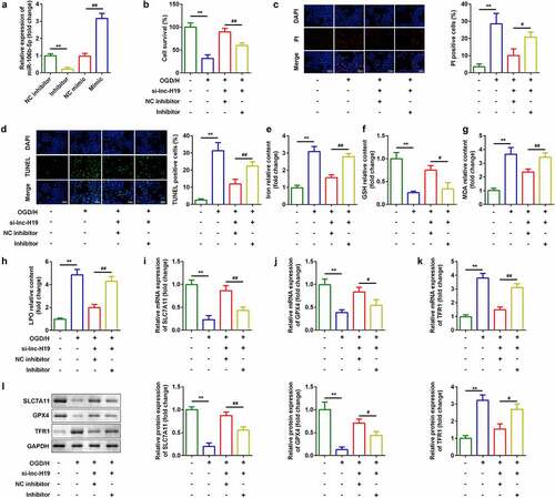
Figure 5. ACSL4 as a target gene of miR-106b-5p
(a) Bioinformatic analysis predicted the binding sites between miR-106b-5p and ACSL4. (b) Dual-luciferase reporter gene assays confirmed that ACSL4 was a target of miR-106b-5p in BMVECs. (c) Interactions involving miR-106b-5p and ACSL4 were determined using RNA pull-down assays. (d) The expression of ACSL4 was determined by RT-qPCR. (e) ACSL4 expression levels in BMVECs with or without treatment by OGD/H.*P < 0.05, **P < 0.01, ##P < 0.01.
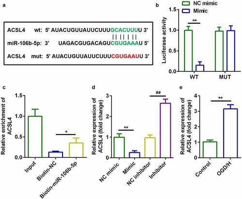
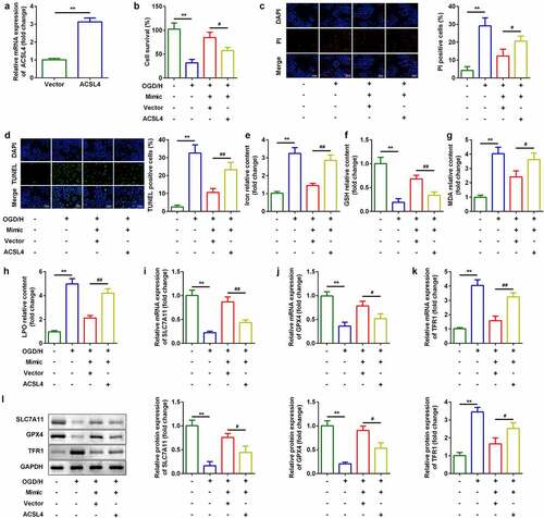
Figure 6. Up-regulation of ACSL4 inhibited the effects of miR-106b-5p
(a) ACSL4 expression was detected using RT-qPCR after miR-106b-5p overexpression. (b) Cell viability was detected using CCK8 assays after transfection with miR-106b-5p mimic. (c) PI staining was used to detect cell death. (d) TUNEL staining was used to detect cell death. (e–h) Levels of iron, GSH, MDA, and LPO. (i–k) Expression of SLC7A11, GPX4, and TFR1 mRNAs were measured using RT-qPCT. (l) Expression of SLC7A11, GPX4, and TFR1 proteins detected by western blotting.**P< 0.01, compared with vector group or control group. #P < 0.05, ##P < 0.01, compared with the OGD/H + mimic + vector group.