Figures & data

Figure 1. EPH attenuate neurological damage in MCAO rats. (a) Rats underwent MCAO and received treatment with EPH (0.5, 1.0 or 1.5 mg/kg) after the onset of ischemia. 72 h post MCAO, neurological function of MCAO rats treated with or without EPH was assessed by neurologic severity scores. N = 20. (b) The number of mistakes in jumping platform of MCAO rats treated with or without EPH was evaluated by step-down test. N = 8. (c) The percentage of spontaneous alternation of MCAO rats treated with or without EPH was assessed by Y maze test. N = 8. (d-e) The expression of BDNF and NGF mRNA and protein was analyzed by western blot. N = 8. *p < 0.05
Figure 2. EPH reduced brain damage in MCAO rats. (a) The volume of cerebral infarction in rats was evaluated by TTC staining. N = 6. (b) The wet and dry method was used to evaluate the brain edema in rats. N = 6. (c) The pathological damage of rat brain tissues was assessed by HE staining. Bar = 20 µm. N = 8. *p < 0.05
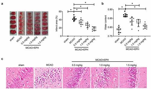
Figure 3. EPH reduced the apoptosis of hippocampal neurons in MCAO rats. (a) The loss and apoptosis of hippocampal neurons in rats were analyzed by Nissl staining and TUNEL assay. Bar = 20 µm. N = 8. (b) The expression of Bax, Bcl-2 and cleaved caspase-3 proteins was analyzed by western blot assay. N = 8. *p < 0.05
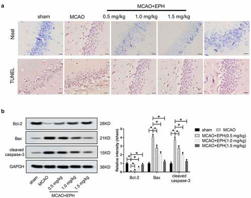
Figure 4. EPH attenuated oxidative stress and inflammation in MCAO rats. (a-d) The levels of MDA, GPx, CAT and NO in brain tissues of rats were analyzed by the corresponding kits. (e-h) The contents of IL-6, TNF-α, IL-4 and IL-10 in brain tissues of rats were detected by ELISA kits. N = 8. *p < 0.05
Figure 5. EPH mitigated brain injury in MCAO rats by activating PI3K/AKT pathway. (a) The expression of p-PI3K, PI3K, p-AKT and AKT in the brain tissues of MCAO rats treated with or without EPH was analyzed by western blot assay. *p < 0.05. Rats received PI3K inhibitor LY294002 intracerebroventricularly to inhibit the PI3K/Akt signaling pathway before MCAO, and EPH (1.5 mg/kg) was injected intraperitoneally into MCAO rats after the onset of ischemia. (b) 24 h after MCAO, the expression of p-PI3K, PI3K, p-AKT and AKT in the brain tissues of MCAO rats treated with EPH or/and LY294002 was analyzed by western blot assay. N = 8. (c) The neurological impairment of MCAO rats treated with EPH or/and LY294002 were analyzed by neurologic severity scores. (d-e) The learning and memory ability of MCAO rats treated with EPH or/and LY294002 were analyzed by step-down test and T-maze test. N = 8. (f) The volume of cerebral infarction in rats was evaluated by TTC staining. (g) Cerebral edema in rats was evaluated by the wet-dry method. N = 6. (h) The brain tissue injury and hippocampal neuronal apoptosis were analyzed by HE staining, Nissl staining and TUNEL staining. Bar = 20 µm. N = 8. (i-j) The expression of BDNF, NGF, Bax, Bcl-2 and cleared caspase-3 proteins was analyzed by western blot assay. N = 8. *p < 0.05
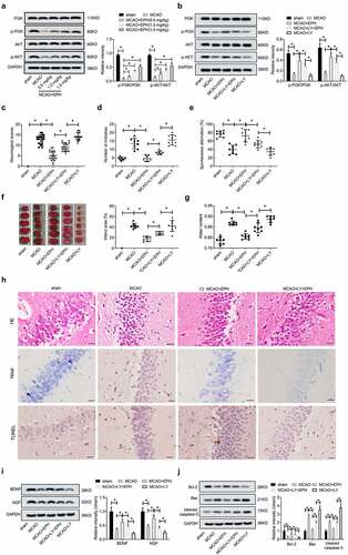
Figure 6. EPH extenuated oxidative stress and inflammatory response in MCAO rats by activating PI3K/AKT pathway. (a-d) The levels of MDA, GPx, CAT and NO in brain tissues of rats were analyzed by the corresponding kits. (e-h) The contents of IL-6, TNF-α, IL-4 and IL-10 in brain tissues of rats were detected by ELISA kits. N = 8. *p < 0.05