Figures & data
Figure 1. Stimulation of LPS affected the proliferation and apoptosis of cervical epithelial cells. (a) CCK-8 was used for the detection of the proliferation of cervical epithelial cells. (b, c) The expression of Ki-67, HMGB1 and RAGE in these cells was detected with the western blotting. (d, e) The apoptosis rates of these cells were determined with the flow cytometry. *p < 0.05, **p < 0.01, ***p < 0.001 vs control
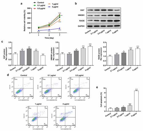
Figure 2. Inhibition of RAGE suppressed the proliferation and migration of cervical epithelial cells. (a) The proliferation of cervical epithelial cells was determined with the CCK-8. (b, c) The cell cycle was detected with the flow cytometry. (d) The expression of Ki-67 and cyclin D1 in cervical epithelial cells was detected with the western blotting. (e, f) The migration of cervical epithelial cells was determined with the wound healing assays. ***p < 0.001 vs control; #p < 0.05, ###p < 0.001 vs LPS
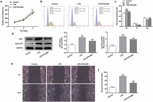
Figure 3. Suppression of RAGE relieved the LPS induced apoptosis of cervical epithelial cells. (a, b) The apoptosis of cervical epithelial cells was determined with the flow cytometry. (c) The expression of apoptosis related proteins was detected with the western blotting. **p < 0.01, ***p < 0.001 vs control; #p < 0.05, ##p < 0.01 vs LPS
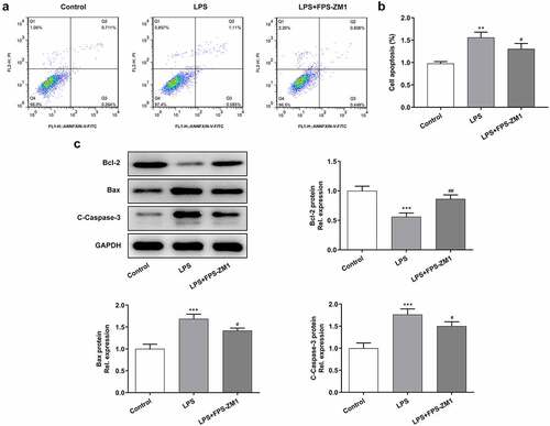
Figure 4. Inhibition of RAGE relieved the LPS induced inflammation of cervical epithelial cells. (a, b, c) The expression of inflammatory factors (IL-1β, IL-6 and TNF-α) was detected with the ELISA. (d) The expression of HMGB1 and RAGE in cervical epithelial cells was detected with the western blotting. (e) The expression of TLR4 and NF-κB p65 in cervical epithelial cells was determined with the western blotting. **p < 0.01, ***p < 0.001 vs control; #p < 0.05, ##p < 0.01, ###p < 0.001 vs LPS
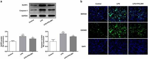
Figure 5. Suppression of RAGE relieved the pyroptosis of cervical epithelial cells. (a) The expression of NLRP3 and caspase4 in cervical epithelial cells was determined with the western blotting. (b) The GSDMD in cervical epithelial cells was observed with the immunofluorescence. ***p < 0.001 vs control; #p < 0.05 vs LPS