Figures & data
Table 1. Primer sequences used for RT-qPCR
Figure 1. LncRNA ZFAS1 expression was downregulated with the increasing concentrations of bupivacaine
(a) MTT assay was used to measure cell viability of SH-SY5Y cells treated with 0, 0.5, 1.0, 1.5, or 2.0 mM bupivacaine. (b) Caspase-3 activity was detected in SH-SY5Y cells treated with different concentrations of bupivacaine. (c) RT-qPCR was performed to detect the expression of ZFAS1 in SH-SY5Y cells treated with different concentrations of bupivacaine. (d) RT-qPCR was used to measure the expression of ZFAS1 in SH-SY5Y cells at 0 h, 6 h, 12 h, 24 h, and 48 h after exposure to 1.0 mM bupivacaine. *P < 0.05.
Figure 2. LncRNA ZFAS1 regulated cell viability and apoptosis in SH-SY5Y cells
(a) The expression of ZFAS1 was detected by RT-qPCR in SH-SY5Y cells transfected with shNC or shZFAS1. (b) MTT assay was used to detect cell viability of bupivacaine-treated SH-SY5Y cells transfected with shNC or shZFAS1. (c) TUNEL assay was utilized to measure cell apoptosis of bupivacaine-treated SH-SY5Y cells transfected with shNC or shZFAS1 (magnification, x200). (d) Caspase-3 activity was measured in bupivacaine-treated SH-SY5Y cells transfected with shNC or shZFAS1. (e) The expression of ZFAS1 was detected by RT-qPCR assay in SH-SY5Y cells transfected with pcDNA3.1 or pcDNA3.1/ZFAS1. (f) MTT assay was used to detect cell viability of bupivacaine-treated SH-SY5Y cells transfected with pcDNA3.1 or pcDNA3.1/ZFAS1. (g) TUNEL assay was utilized to measure cell apoptosis of bupivacaine-treated SH-SY5Y cells transfected with pcDNA3.1 or pcDNA3.1/ZFAS1 (magnification, x200). (h) Caspase-3 activity was measured in bupivacaine-treated SH-SY5Y cells transfected with pcDNA3.1 or pcDNA3.1/ZFAS1. **P < 0.01.
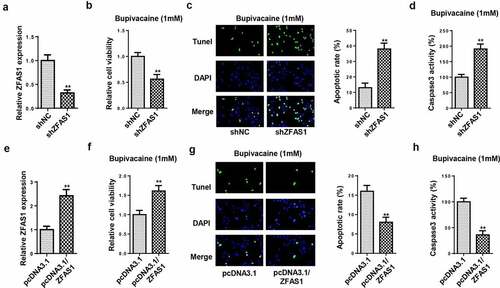
Figure 3. ZFAS1 directly interacted with miR-421
(a) The predicted targeting sequence of miR-421 on the ZFAS1. (b) RT-qPCR was performed to detect the expression of miR-421 in SH-SY5Y cells treated with 0, 0.5, 1.0, 1.5, or 2.0 mM bupivacaine. (c) RT-qPCR was used to measure the expression of miR-421 in SH-SY5Y cells at 0 h, 6 h, 12 h, 24 h, and 48 h after exposure to 1.0 mM bupivacaine. (d and e) Dual-luciferase reporter and RIP assays were utilized to confirm the interaction between ZFAS1 and miR-421. (f) The expression of miR-421 was detected by RT-qPCR in SH-SY5Y cells transfected with pcDNA3.1, pcDNA3.1-ZFAS1, shNC, or shZFAS1. *P < 0.05, **P < 0.01, ***P < 0.001.
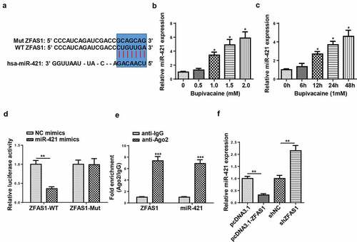
Figure 4. ZFAS1 mitigated bupivacaine-induced neurotoxicity via miR-421
(a) RT-qPCR was used to measure the expression of miR-421 in SH-SY5Y cells transfected with shNC, shZFAS1, or shZFAS1+ miR-421 inhibitor. (b) MTT assay was used to detect cell viability of bupivacaine-treated SH-SY5Y cells transfected with shNC, shZFAS1, or shZFAS1+ miR-421 inhibitor. (c) TUNEL assay was utilized to detect cell apoptosis of bupivacaine-treated SH-SY5Y cells transfected with shNC, shZFAS1, or shZFAS1+ miR-421 inhibitor (magnification, x200). (d) Caspase-3 activity was detected in bupivacaine-treated SH-SY5Y cells transfected with shNC, shZFAS1, or shZFAS1+ miR-421 inhibitor. *P < 0.05, **P < 0.01.
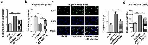
Figure 5. ZNF564 was the direct target of miR-421
(a) The potential downstream targets were predicted using miRmap and RNA22 database. (b) The expression of ZNF564 was detected in SH-SY5Y cells treated with 0, 0.5, 1.0, 1.5, or 2.0 mM bupivacaine by RT-qPCR. (c) The expression of ZNF564 was detected by RT-qPCR in SH-SY5Y cells at 0 h, 6 h, 12 h, 24 h, and 48 h after exposure to 1.0 mM bupivacaine. (d) The predicted binding site between miR-421 and ZNF564. (e) Luciferase activity was detected in SH-SY5Y cells co-transfected with ZNF564-WT or ZNF564-Mut and miR-421 mimics or NC mimics. (f) The expression of ZNF564 was detected by RT-qPCR in SH-SY5Y cells transfected with miR-421 mimics or miR-421 inhibitor. *P < 0.05, **P < 0.01.
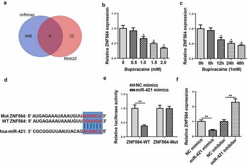
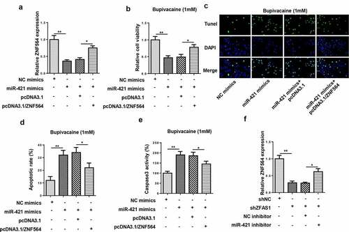
Figure 6. ZFAS1 regulated bupivacaine-induced neurotoxicity via the miR-421/ZNF564 axis
(a) The expression of ZNF564 was detected by RT-qPCR in SH-SY5Y cells transfected with NC inhibitor, miR-421 inhibitor, miR-421 inhibitor+shNC, or miR-421 inhibitor+shZNF564. (b) MTT assay was performed to detect cell viability of bupivacaine-treated SH-SY5Y cells transfected with NC inhibitor, miR-421 inhibitor, miR-421 inhibitor+shNC, or miR-421 inhibitor+shZNF564. (c and d) TUNEL assay was used to measure the apoptosis rate of bupivacaine-treated SH-SY5Y cells transfected with NC inhibitor, miR-421 inhibitor, miR-421 inhibitor+shNC, or miR-421 inhibitor+shZNF564 (magnification, x200). (e) Caspase-3 activity was detected in bupivacaine-treated SH-SY5Y cells transfected with NC inhibitor, miR-421 inhibitor, miR-421 inhibitor+shNC, or miR-421 inhibitor+shZNF564. (f) The expression of ZNF564 was detected in SH-SY5Y cells. *P < 0.05, **P < 0.01.