Figures & data
Figure 1. PEAK1 plays a critical role in migration and invasion in melanoma cells in vitro
A, PEAK1 protein expression was detected by western blot assay in SKMEL-1, SKMEL-2, SKMEL-19 and SKMEL-28 cells. B, PEAK1 protein was detected by western blot assay in SKMEL-19 cells transfected with PEAK1 or control for 48 h. C, PEAK1 protein was detected by western blot assay in SKMEL-28 cells transfected with PEAK1 shRNA or control NC shRNA for 48 h. D, E, transwell migration assay and transwell invasion assay in PEAK1 or control transfected SKMEL-19 cells. F, G, transwell migration and invasion assay in PEAK1 shRNA or control NC shRNA transfected SKMEL-28 cells.
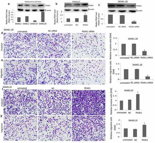
Figure 2. PEAK1 regulates SKMEL19/28 cells migration in vitro by wound healing assay
SKMEL19 cells were transfected with PEAK1 or NC, SKMEL28 cells were transfected with PEAK1 shRNA or NC shRNA. Cells were then grown in a monolayer, wounded and left to recover the wound then imaged at the same frame after 48 h. The graph is a quantitation of the wounds. Wound widths were measured at 11 different points for each condition and the average rate of wound closure was calculated in μm/hr. Data are the mean −/+ SEM from 3 wound closure assays and *p < 0.01. Scale bar is 200 μm.
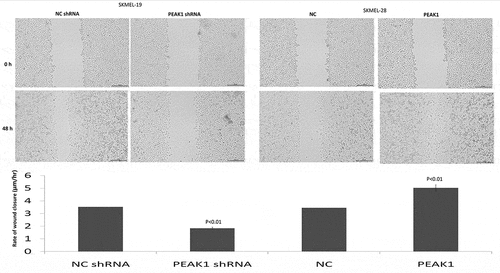
Figure 3. PEAK1- induced cell invasion and migration by regulating JAK/STAT3 signals
A, JAK/STAT3 signals were detected in NC or PEAK1 transfected SKMEL-19 cells by western blot assay. B, JAK/STAT3 signals were detected in NC shRNA or PEAK1 shRNA transfected SKMEL-28 cells by western blot assay. C, SKMEL-19 cells were treated with P6, then transfected with NC or PEAK1 for 48 h, cell invasion was detected by transwell invasion assay. D, SKMEL-19 cells were treated with P6, then transfected with NC or PEAK1 for 48 h, cell invasion was detected by transwell migration assay.
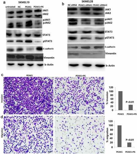
Figure 4. PEAK1 plays a critical role in lung metastasis in melanoma cells in vivo
A, SKMEL19/PEAK1 or NC cells were i.v tail vein injected into nude mice for 35 days. Lung metastatic nodes were counted in the lungs. B, SKMEL28 /Lv-NC shRNA or Lv-PEAK1 shRNA cells were i.v tail vein injected into nude mice for 35 days. Lung metastatic nodes were counted in the lungs. C, E-cadherin expression was detected in metastatic lung nodes of PEAK1 or NC transfected mices. D, E-cadherin expression was detected in metastatic lung nodes of Lv-NC shRNA or Lv-PEAK1 shRNA transfected mices. E, vimentin expression was detected in metastatic lung nodes of PEAK1 or NC transfected mices. F, vimentin expression was detected in metastatic lung nodes of Lv-NC shRNA or Lv-PEAK1 shRNA transfected mice.
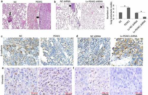
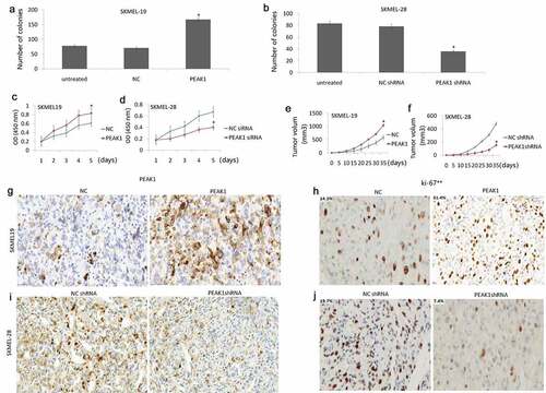
Figure 5. PEAK1 plays a critical role in melanoma cell growth in vitro and in vivo.
Cell growth was detected by colony formation assay in SKMEL19/PEAK1 or NC cells (A) and SKMEL28/Lv-NC shRNA or Lv-PEAK1 shRNA cells (B). Cell proliferation assay by CCK-8 assay in SKMEL19/PEAK1 or NC cells (C) and SKMEL28/Lv-NC shRNA or Lv-PEAK1 shRNA cells (D). SKMEL-19/PEAK1 or NC (E) or SKMEL28/Lv-NC shRNA or Lv-PEAK1 shRNA cells (F) were inoculated subcutaneously into nude mice. Xenografts were measured every 5 days with a caliper. PEAK1 (G) and Ki-67 (H) expression was detected in SKMEL19 Xenografts. PEAK1 (I) and Ki-67 (J) expression was detected in SKMEL28 Xenografts.