Figures & data
Table 1. List of primers used in this study
Figure 1. MEX3A expression is increased in breast cancer. (a) MEX3A expression in breast cancer based on GEPIA database. (b) Immunohistochemistry was used to measure MEX3A expression levels in clinical specimens. (c) The strong positive expression rate, weak positive expression rate and negative expression rate of MEX3A in clinical samples. (d) MEX3A mRNA expression level in clinical samples. Western blotting (e) and RT-qPCR (f) were used to measure MEX3A expression in breast cancer cells and MCF-10A cells. *P < 0.05
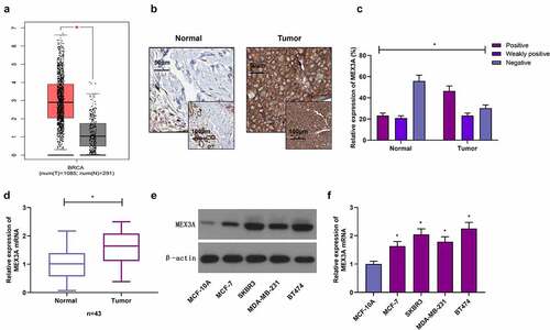
Figure 2. High MEX3A expression is associated with the poor prognosis of breast cancer patients. (a) The relationship between MEX3A (probe: 226346) and the overall survival of breast cancer patients. (b) The relationship between the expression level of MEX3A (probe: 236885) and the overall survival of breast cancer patients
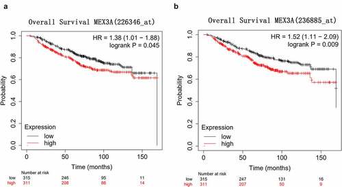
Figure 3. MEX3A promotes the proliferation and migration of breast cancer cells. RT-qPCR (a) and Western blotting (b-c) to measure MEX3A expression levels. (d) CCK-8 assay to evaluate cell proliferation. (e-h) The wound healing assay and Transwell assay to assess cell migration and invasion respectively. *P < 0.05
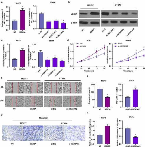
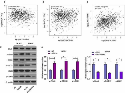
Figure 4. MEX3A activates RhoA/ROCK1/LIMK1 signaling in breast carcinoma cells. (a-c) The relationship between the expression levels of RhoA, ROCK1 and LIMK1 proteins and MEX3A expression level by using GEPIA database. (d and e) The protein expression of p-RhoA, p-ROCK1 and p-LIMK1 in breast cancer cells with MEX3A overexpressed or knocked down. *P < 0.05