
Figures & data
Figure 1. circUSP36 expression was upregulated in ox-LDL-treated endothelial cells. (a) qRT-PCR analysis for circUSP36 expression in endothelial cells treated with or without RNase R digestion (n = 3). (b) qRT-PCR detection of circUSP36 and linear mRNA in endothelial cells exposed to Actinomycin D (n = 3). (c) Divergent primers could amplify circUSP36 in cDNA but not gDNA (n = 3). (d) circUSP36 expression in endothelial cells treated with or without ox-LDL was detected via use of qRT-PCR assay (n = 3). **P < 0.01
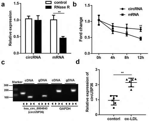
Figure 2. Ectopic expression of circUSP36 attenuated endothelial cell proliferation and migration. (a) The efficiency of circUSP36 overexpression was examined by qRT-PCR (n = 3). (b) CKK-8 assay explored the viability of ox-LDL-treated endothelial cells in different groups (n = 6). (c-e) Transwell and wound-healing assays measured the migration ability of endothelial cells exposed to ox-LDL for the indicated times after transfection (n = 6). Cells migrating to the underside of the transwell insert were counted. **P < 0.01
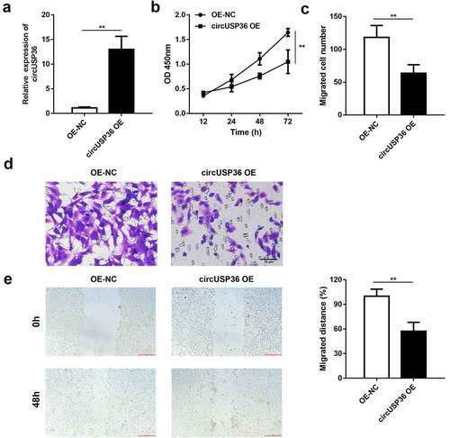
Figure 3. circUSP36 adsorbed miR-637 in a sponge form. (a) Binding sites between circUSP36 and miR-637 as predicted by circInteractome. (b) Interaction between circUSP36 and miR-637 was evaluated with a luciferase reporter assay (n = 3). (c) Effects of circUSP36 on miR-637 expression were assessed by qRT-PCR (n = 3). (d) RNA pull-down assay tested the binding affinity between circUSP36 and miR-637 (n = 3). (e) FISH was conducted for the co-localization of circUSP36 and miR-637 in endothelial cells (n = 3). **P < 0.01
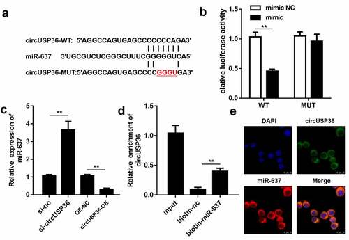
Figure 4. Overexpression of miR-637 relieved the role of circUSP36 in endothelial cells. (a) Overexpression efficacy of miR-637 mimic was assessed by qRT-PCR (n = 3). (b) Cell proliferation in four different groups was examined with the CCK-8 assay (n = 6). (c-f) Cell migration in four different groups was estimated by transwell and wound-healing assays (n = 6). Cells migrating to the underside of the transwell insert were counted. **P < 0.01
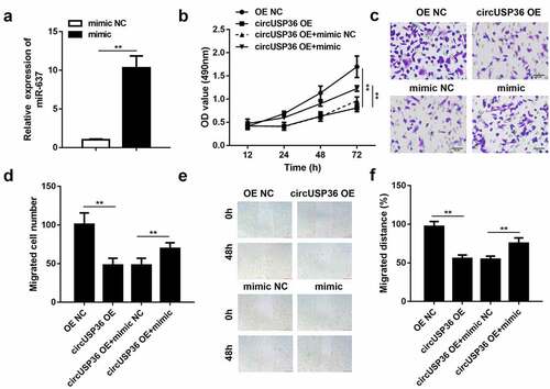
Figure 5. WNT4 functioned as a downstream target of miR-637. (a) Binding sequences of miR-637 in WNT4 3ʹ UTR. (b) Luciferase reporter assay was employed to validate the targeting relationship between miR-637 and WNT4 (n = 3). (c) qRT-PCR and western blotting detection of WNT4 expression in endothelial cells transfected with miR-637 mimic/inhibitor or their negative controls (n = 3). (d) Interplay between miR-637 and WNT4 was estimated by the RNA pull-down assay (n = 3). **P < 0.01
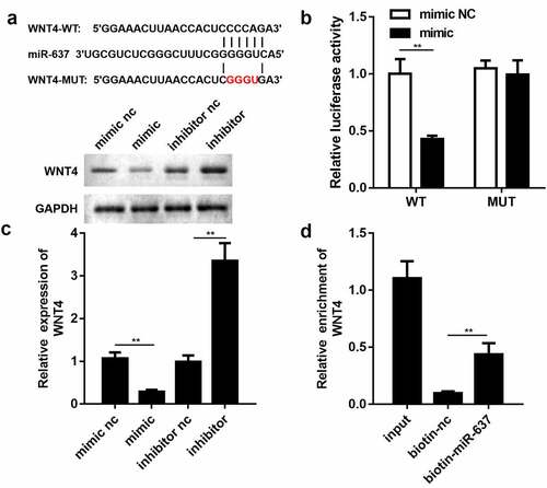
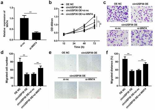
Figure 6. WNT4 was responsible for effects of circUSP36 on endothelial cell behavior. (a) qRT-PCR detection of WNT4 expression in endothelial cells following different treatments (n = 3). (b) CCK-8 analysis for testing cell proliferation in different groups (n = 6). (c-f) Ability of ox-LDL-treated endothelial cells to migrate was assessed by transwell and wound-healing experiments. (n = 6) Cells migrating to the underside of the transwell insert were counted. **P < 0.01
Data Availability
All the data is available from the corresponding author due to reasonable request