
Figures & data
Figure 1. Elevated CDCA7 expression in tumor tissues of OC patients
(a) Expression levels of CDCA7 in OC tissues and adjacent normal tissues were obtained at Gene Expression Profiling Interactive Analysis (GEPIA; http://gepia.cancer-pku.cn/index.html). (b) IHC staining for determination of CDCA7 expression in 5 pairs of clinical tumor tissue specimens and adjacent normal ones. (c) Western blot assay for determination of CDCA7 protein level in 5 pairs of clinical tumor tissue specimens and adjacent normal ones. (d) RT-qPCR for determination of CDCA7 mRNA level in five pairs of clinical tumor tissue specimens and adjacent normal ones. *p < 0.05, **p < 0.01, ***p < 0.001.
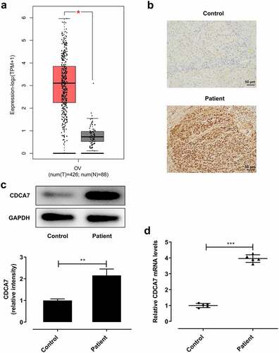
Figure 2. Elevated CDCA7 expression in OC cell lines
(a) Expression levels of CDCA7 in various cancer cell lines were obtained at Cancer Cell Line Encyclopedia (CCLE; https://portals.broadinstitute.org/ccle/) database. (b) Western blot assay for determination of CDCA7 protein level in normal ovarian epithelial cells (NOEC) and ovarian cancer cells (OVCAR3, SKOV3, CAOV-3, A2780). (c) RT-qPCR for determination of CDCA7 mRNA level in normal ovarian epithelial cells (NOEC) and ovarian cancer cells (OVCAR3, SKOV3, CAOV-3, A2780). **p < 0.01, ***p < 0.001.
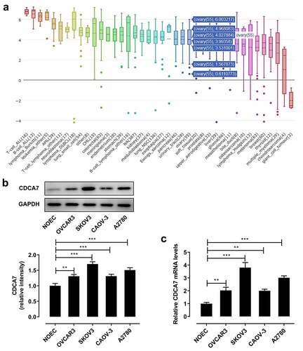
Figure 3. CDCA7 silencing arrested OC progression
(a) Western blot assay was applied to validate the transfection efficiency in SKOV3 cells following introduction of shRNA-CDCA7-1 or shRNA-CDCA7-2. (b) RT-qPCR was applied to validate the transfection efficiency in SKOV3 cells following introduction of shRNA-CDCA7-1 or shRNA-CDCA7-2. (c) CCK-8 assay for determination of OC cell proliferation. (d) Flow cytometry analysis for determination of OC cell cycle. (e) Quantitative analysis of cell cycle distribution of SKOV3 cells. (f) Western blot assay for determination of CyclinE1 and CyclinE2 protein levels. *p < 0.05, **p < 0.01, ***p < 0.001.
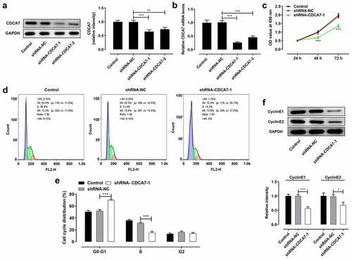
Figure 4. CDCA7 silencing restrained the migrative and invasive abilities of OC cells
(a) Wound healing assay for determination of the migration of SKOV3 cells. (b) Transwell assay for determination of the invasion of SKOV3 cells. (c) Western blot assay for determination of MMP2 and MMP9 protein levels. **p < 0.01, ***p < 0.001.
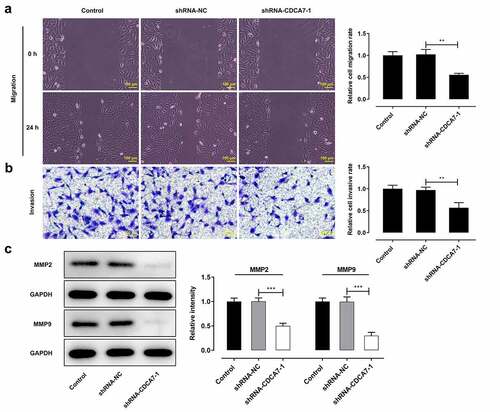
Figure 5. CDCA7 silencing repressed in vitro angiogenesis of HUVECs
a) Western blot assay for determination of VEGFA, VEGFR1, and VEGFR2 protein levels. (b) Tube formation assay of HUVECs. (c) Quantitative analysis of the angiogenesis ability. **p < 0.01, ***p < 0.001.
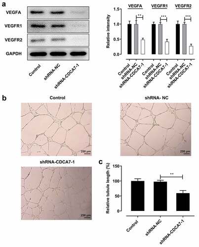
Figure 6. CDCA7 interacted with EZH2
(a) GeneMANIA database analysis of the interaction between CDCA7 and EZH2. (b, c) Co-IP assay of the interaction between CDCA7 and EZH2. (d, e) SKOV3 cells were introduced with shRNA-CDCA7. Western blot assay and RT-qPCR were applied to assess the regulating effects of CDCA7 knockdown on EZH2 protein and mRNA levels. (f, g) SKOV3 cells were introduced with EZH2 overexpression plasmid. Western blot assay and RT-qPCR were applied to evaluate the transfection efficiency. (h, i) SKOV3 cells were transfected with shRNA-CDCA7 or cotransfected with shRNA-CDCA7 and EZH2 overexpression plasmid. Western blot assay and RT-qPCR were applied to assess the influence of EZH2 overexpression plasmid on the regulating effects of CDCA7 knockdown. **p < 0.01, ***p < 0.001.
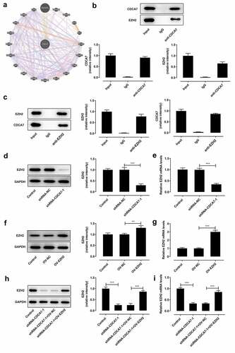
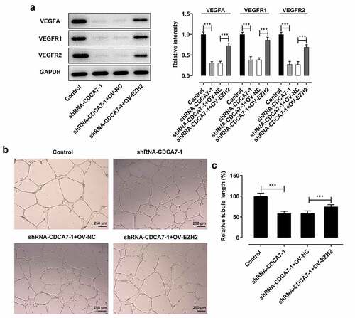
Figure 7. CDCA7 silencing arrested angiogenesis by suppressing EZH2 expression
(a) Western blot assay for determination of VEGFA, VEGFR1, and VEGFR2 protein levels. (b) Tube formation assay of HUVECs. (c) Quantitative analysis of the angiogenesis ability. ***p < 0.001.
Availability of data and materials
The datasets used and/or analyzed during the present study are available from the corresponding author on reasonable request.