Figures & data
Table 1. RT-qPCR primer sequences
Figure 1. Elevated circSMEK1 expression in NP
A-B. Mechanical pain threshold and withdrawal latency to thermal stimulation in plantar test of Sham and CCI groups; C. Immunohistochemistry detection of Iba1 expression in spinal cord tissue of Sham and CCI groups; D. Flow cytometry detection of CD68+ and CD206+ cells in spinal cord tissues of Sham and CCI groups; E. ELISA detection of TNF-α, IL-1β and IL-6 levels in the spinal cord tissues of Sham and CCI groups; F. ELISA detection of TNF-α, IL-1β and IL-6 levels in microglia of Control and LPS groups; G. Western blot detection of CD11b, CD68 and CD206 expressions in spinal cord of Sham and CCI groups; H. Western blot detection of CD11b, CD68 and CD206 expression in microglia of Control and LPS groups; I. RT-qPCR detection of circSMEK1 expression in spinal cord tissue of Sham and CCI groups; J. RT-qPCR detection of circSMEK1 expression in microglia of Control and LPS groups; Data presentation was detailed as mean ± SD (A, B, D, F, H, n = 9; E, G, I, n = 3), * vs Sham or Control groups, P < 0.05.
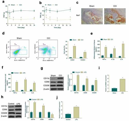
Figure 2. CircSMEK1 knockdown improves NP, while overexpressed circSMEK1 aggravates it
A. RT-qPCR detection of circSMEK1 expression in spinal cord tissue of LV-NC and LV-circSMEK1 groups; B-C. Mechanical pain threshold and heat-stimulated withdrawal latency in plantar test of LV-NC and LV-circSMEK1 groups; D. Immunohistochemistry detection of Iba1 expression in spinal cord of LV-NC and LV-circSMEK1 groups; E. Flow cytometry detection of CD68+ and CD206+ cells in spinal cord tissues of rats in LV-NC and LV-circSMEK1 groups; F. ELISA detection of TNF-α, IL-1β and IL-6 levels in spinal cord tissue of LV-NC and LV-circSMEK1 groups; G. Western blot detection of CD11b, CD68 and CD206 expressions in spinal cord tissue of LV-NC group and LV-circSMEK1 group; H. RT-qPCR detection of CD11b, CD68 and CD206 expressions in microglia of pcDNA3.1 and pcDNA3.1-CircSMEK1 groups; I. ELISA detection of TNF-α, IL-1β and IL-6 levels in microglia of pcDNA3.1 and pcDNA3.1-circSMEK1 groups; J. Western blot detection of CD11b, CD68 and CD206 expressions in microglia of pcDNA3.1 and pcDNA3.1-circSMEK1 groups; Data presentation detailed as mean ± SD (A-F, n = 9; G-I, n = 3); * vs LV-NC or pcDNA 3.1 groups, P < 0.05.
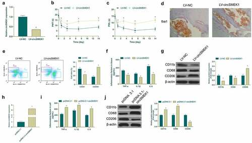
Figure 3. Knocking down miR-216a-5p promotes NP, but overexpressed miR-216a-5p represses it
A. RT-qPCR detection of miR-216a-5p expression in CCI rats and LPS-treated microglia; B. RT-qPCR detection of miR-216a-5p expression in spinal cord of LV-NC and LV-miR-216a-5p groups; C-D. Mechanical pain threshold and heat-stimulated withdrawal latency of thermal stimulation in plantar test of LV-NC and LV-miR-216a-5p groups; E. Immunohistochemistry detection of Iba1 expression in spinal cord of LV-NC and LV-miR-216a-5p groups; F. Flow cytometry detection of CD68+ and CD206+ cells in spinal cord tissues of rats in LV-NC and LV- miR-216a-5p groups; G. ELISA detection of TNF-α, IL-1β and IL-6 levels in spinal cord of LV-NC and LV-miR-216a-5p groups; H. Western blot detection of CD11b, CD68 and CD206 expressions in spinal cord tissue of LV-NC and LV-miR-216a-5p groups; I. RT-qPCR detection of circSMEK1 expression in microglia of mimic NC and miR-216a-5p-mimic groups; J. ELISA detection of TNF-α, IL-1β and IL-6 levels in microglia in mimic NC and miR-216a-5p-mimic groups; K. Western blot detection of CD11b, CD68 and CD206 expressions in microglia of mimic NC and miR-216a-5p-mimic groups; Data presentation detailed as mean ± SD (A, n = 9 or 3; B-G, n = 9; H-J, n = 3); * vs LV-NC or mimic NC groups, P < 0.05.
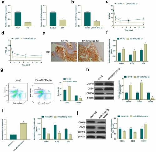
Figure 4. circSMEK1 competitively binds to miR-216a-5p
A. RT-qPCR detection of miR-216a-5p expression in LV-NC and LV-circSMEK1 groups; B. RT-qPCR detection of miR-216a-5p expression in pcDNA3.1 and pcDNA3.1-circSMEK1 groups; C. Queried the potential binding sites of circSMEK1 and miR-216a-5p through http://starbase.sysu.edu.cn/; D; Detection of targeting relationship between circSMEK1 and miR-216a-5p via DLR experiment; Data presentation detailed as mean ± SD (A, n = 9; B and D, n = 3), * vs LV-NC, pcDNA3.1, or mimic NC groups, P < 0.05.
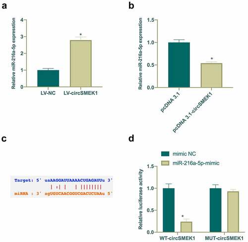
Figure 5. TXNIP is targeted via miR-216a-5p
A. Western blot detection of TXNIP expression of LV-NC and LV-miR-216a-5p groups; B. Western blot detection of TXNIP expression in microglia of mimic NC and miR-216a-5p-mimic groups; C. Checked the potential-binding sites of TXNIP and miR-216a-5p via website http://starbase.sysu.edu.cn/; D; DLR experiment detection of the targeting relationship of TXNIP and miR-216a-5p; Data presentation detailed as mean ± SD (A, n = 9; B and D, n = 3); * vs LV-NC or mimic NC groups, P < 0.05.
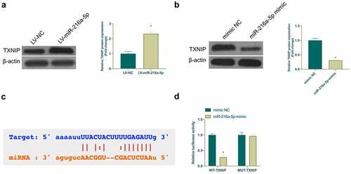
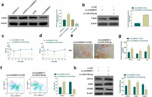
Figure 6. circSMEK1 promotes NP through the miR-216a-5p/TXNIP axis
In LV-circSMEK1+ LV-NC and LV-circSMEK1+ LV-miR-216a-5p groups, A. Western blot detection of the effect of knockdown or overexpression of circSMEK1 on TXNIP expression in microglia; B. Western blot detection of TXNIP expression in rat spinal cord tissues; C and D. Mechanical pain threshold and withdrawal latency to thermal stimulation in plantar test; E. Immunohistochemistry detection of Iba1 expression in spinal cord tissue; F. Flow cytometry detection of the ratio of CD68+ and CD206+ cells in rat spinal cord; G. ELISA detection of TNF-α, IL-1β and IL-6 levels in spinal cord tissue; H. Western blot detection of CD11b, CD68 and CD206 expressions in spinal cord tissue; Data presentation detailed as mean ± SD (n = 9); * vs LV-circSMEK1 + LV-NC group, P < 0.05.