Figures & data
Figure 1. CTRP6 relieved the sevoflurane induced injury of central nervous tissues. (a) H&E staining was performed for the detection of the injury of central nervous tissues. (b) Immunofluorescence assay was performed to detect the expression of CTRP6 and NeuN in these tissues. #p< 0.05, **p< 0.01
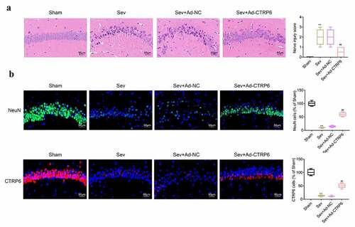
Figure 2. CTRP6 alleviated the sevoflurane induced apoptosis of central nervous tissues. (a) Apoptosis of central nervous tissues was detected with the tunel staining. (b) Western blotting was performed to measure the expression of apoptosis related proteins in central nervous tissues. #p< 0.05, **p< 0.01
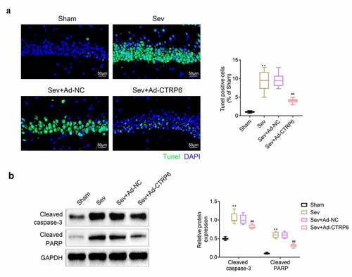
Figure 3. CTRP6 recovered the learning and cognitive abilities of mice. (a) The swimming path of mice were recorded. (b) The length of swimming path was recorded with the computer. (c) The escape latency of these mice in the pool was recorded. (d) Swimming speed of these mice was recorded. (e) Time spend in the platform quadrant of these mice was recorded. (f) Number of platform crossings of these mice was recorded with the computer. #p< 0.05, **p< 0.01
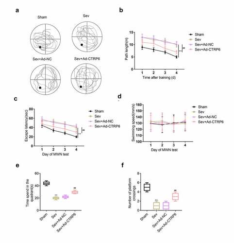
Figure 4. Overexpression of CTRP6 relieved the sevoflurane induced apoptosis of primary cells of central nervous tissue. (a) CCK-8 assays were performed to measure the viability of the primary cells. (b) Flow cytometry was performed to determine the apoptosis of these cells. (c) Western blotting was applied for measuring the expression of apoptosis related proteins in these primary cells. #p< 0.05, **p< 0.01
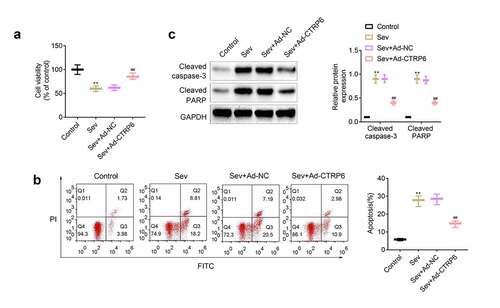
Figure 5. Overexpression of CTRP6 alleviated the sevoflurane induced oxidative stress of primary cells of central nervous tissue. (a) Probe was used for the detection of the ROS levels in these primary cells. (b, c) Commercial kits were used for the measurement of the levels of MDA and SOD in these primary cells. #p< 0.05, **p< 0.01
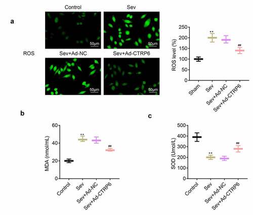
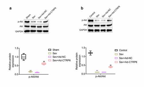
Figure 6. CTRP6 activated the expression of p-Akt in primary cells of central nervous tissue. (a, b) The expression levels of p-Akt and Akt in central nervous tissues and primary cells were measured with western blotting. #p< 0.05, **p< 0.01