Figures & data
Table 1. Primer sequences
Figure 1. TTK overexpression is significantly up-regulated in OC, and is associated with the prognosis of OC patients
A. Venn diagrams illustrating the number of up-regulated differentially expressed genes (DEGs) in three gene expression datasets (GSE14407, GSE18520 and GSE36668). B. GEPIA2 database was used to analyze TTK expression in OC tissues (red), compared with normal ovarian tissues (gray). C. TTK expression in OC tissues and normal tissues was detected by qRT-PCR. D. TTK expression in OC cell lines (SKOV-3, OVCAR-3 and H8910) and normal ovarian epithelial cell lines (HOSE) was detected by qRT-PCR. E. IHC staining was employed to detect TTK protein expression in OC tissue and paracancerous tissue. F. Kaplan-Meier plots were utilized to analyze the relationship between TTK expression and overall survival of OC patients. G. The association between TTK expression and FIGO stage of OC patients. H. The association between TTK expression with differentiation grade of OC patients. * P < 0.05, and *** P < 0.001.
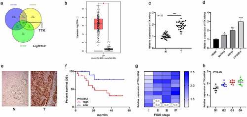
Figure 2. TTK enhances OC cell growth and migration and inhibits apoptosis
A. The pcDNA3.1-TTK and si-TTK#1/si-TTK#2 were transfected into SKOV-3 and OVCAR-3 cell lines, respectively, and the transfection efficiency was examined by qRT-PCR. B. CCK-8 assay was utilized to detect the growth of OC cells. C. Migration assay was executed to detect the migration of OC cells. D. Flow cytometry was performed to detect the apoptosis rate of transfected OC cells. *P < 0.05, **P < 0.01, ***P < 0.001.
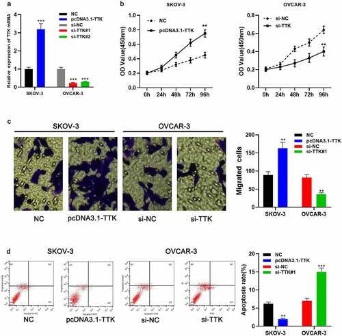
Figure 3. TTK is a downstream target of miR-582-5p
A. The binding site of miR-582-5p and TTK was predicted by bioinformatics analysis. B. Dual-luciferase reporter gene assay was used to validate the binding site between miR-582-5p and TTK. C. qRT-PCR and Western blot experiments were executed to detect the effects of miR-582-5p mimics and inhibitors on TTK expression. D. qRT-PCR was adopted to detect miR-582-5p expression in OC tissues. E. Pearson correlation coefficient analysis was applied to measure the correlation between miR-582-5p and TTK expression in OC tissues. *** P < 0.001.
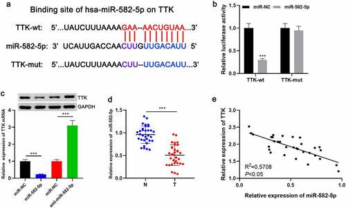
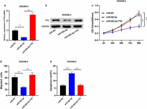
Figure 4. MiR-582-5p restrains the malignant phenotypes of OC cells via regulating TTK
A-B. OVCAR-3 cells were transfected with miR-NC, miR-582-5p mimics, and miR-582-5pmimics + pcDNA3.1-TTK, respectively, and TTK expression in OVCAR-3 cells was detected by qRT-PCR and Western blot assays. C. CCK-8 assay was conducted to detect the growth of OC cells after the transfection. D. Transwell assay was performed to detect the migration of OC cells after the transfection. E. Flow cytometry was carried out to detect the apoptosis rate of transfected OC cells. * P < 0.05, ** P < 0.01, and *** P < 0.001.