Figures & data
Table 1. Primers for qPCR
Figure 1. The expression of COL4A2-AS1 of the colorectal cancer tissues and cell lines were increased. (a): RT-qPCR showed that the mRNA level of COL4A2-AS1 of colorectal cancer tissues was improved. (b): RT-qPCR showed that the mRNA level of COL4A2-AS1 of colorectal cancer cell lines was improved. (c): RT-qPCR showed that the mRNA level of HIF1A of colorectal cancer tissues was improved. D: RT-qPCR showed that the mRNA level of miR-20b-5p of colorectal cancer tissues was decreased. ***P < 0.001, **P < 0.01, *P < 0.05
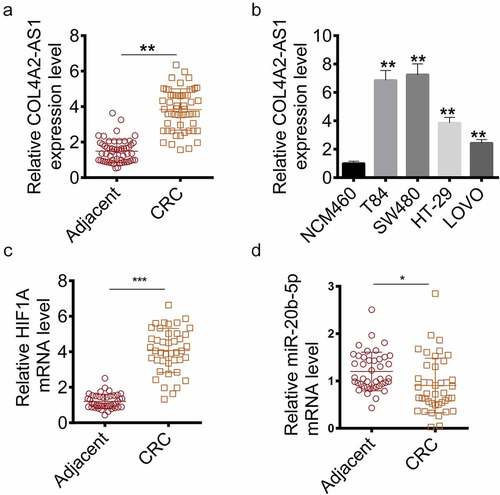
Figure 2. The viability and proliferation of colorectal cancer cells were inhibited by Knockdown of COL4A2-AS1. (a): COL4A2-AS1 expression was overexpressed by pcDNA3.1- COL4A2-AS1. (b): COL4A2-AS1 expression was silenced by shCOL4A2-AS1. (c–d): CCK-8 assay showed that viabilities of T84 and SW480 cells were increased by COL4A2-AS1. (e–f): CCK-8 assay showed that viabilities of T84 and SW480 cells were suppressed by shCOL4A2-AS1. (g): Apoptosis T84 and SW480 cells was improved by shCOL4A2-AS1. (h): Clone populations of T84 and SW480 cells were inhibited by shCOL4A2-AS1. (i): Clone populations of T84 and SW480 cells were increased by COL4A2-AS1. *P < 0.05, **P < 0.01
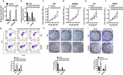
Figure 3. Knockdown of COL4A2-AS1 inhibited tumorigenesis of colorectal cancer in vivo. (a): Tumor growth was inhibited in the shCOL4A2-AS1 mouse compared with the control. (b): HE staining and Ki-67 staining. (c): Tumor size (mm3). (d): Tumor weight (g). €: The expressions level of COL4A2-AS1 in the tumor tissues. **P < 0.01
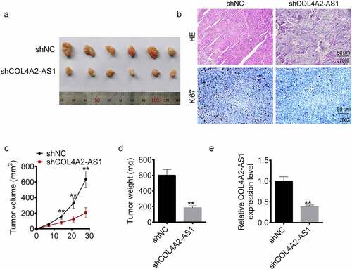
Figure 4. Down-regulation of COL4A2-AS1 expression inhibited aerobic glycolysis. (a–c): Overexpressed COL4A2-AS1 promoted lactate production, glucose uptake and intracellular ATP content of T84 and SW480 cells. (d–f): Downregulated expression of COL4A2-AS1 inhibited lactate production, glucose uptake and intracellular ATP content of T84 and SW480 cells. (g): Overexpressed COL4A2-AS1 decreased the protein expressions of PKM2, GLUT1, LDGA and HK2 of T84 and SW480 cells, while downregulation of COL4A2-AS1 produced opposite effects. **P < 0.01, ##P < 0.01
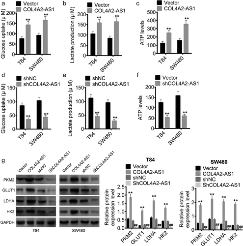
Figure 5. COL4A2-AS1 bound to miR-20b-5p and down-regulated miR-20b-5p expression. (a): LncACTdb predicted the binding region of miR-20b-5p and COL4A2-AS1. The effects of wt-COL4A2-AS1 or mut-COL4A2-AS1 on miR-20b-5p expression were detected by performing dual-luciferase reporter assay on T84 (b) and SW480 cells (c). (d): The expression level of miR-20b-5p was increased by miR-20b-5p mimic. (e–h): RIP experiments were performed on T84 and SW480 cells with Ago2 antibody, and the co-precipitated RNA was subjected to qPCR for COL4A2-AS1 and miR-20b-5p. **P < 0.01
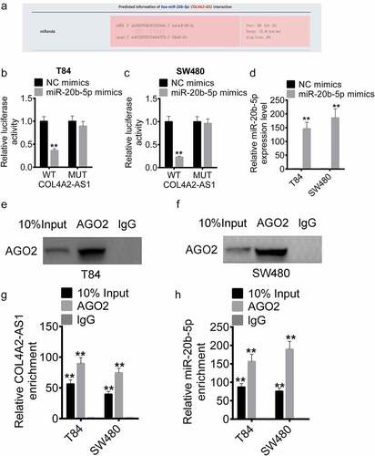
Figure 6. COL4A2-AS1 acted as a ceRNA of miR-20b-5p to regulate HIF1A expression. (a): TargetScan predicted the binding region between miR-20b-5p and the HIF1A 3ʹ-UTR. The effects of wt-HIF1A or mut-HIF1A on miR-20b-5p expression were detected by dual-luciferase reporter assay on T84 (c) and SW480 cells (d). (d): The expression of miR-20b-5p was decreased by miR-20b-5p inhibitor. (e): Western blot was performed for determining the expressions of HIF1A in T84 and SW480 cells transfected with control, COL4A2-AS1, miR-20b-5p mimics or COL4A2-AS1 + miR-20b-5p mimic. (f): Western blot was performed for determining the expression of HIF1A in T84 and SW480 cells transfected with control, shCOL4A2-AS1, miR-20b-5p inhibitor or shCOL4A2-AS1 + miR-20b-5p inhibitor. **P < 0.01, ##P < 0.01
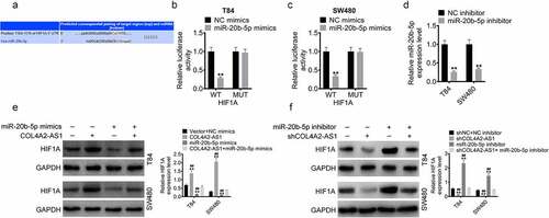
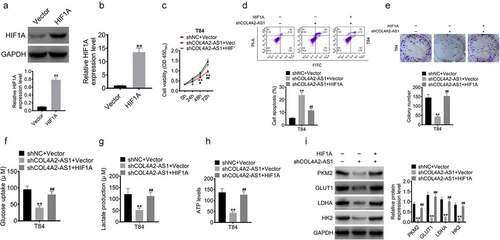
Figure 7. HIF1A overexpression reversed the inhibition of cell proliferation and glycolytic ability induced by knockdown of COL4A2-AS1. (a–b): The protein and mRNA levels of HIF1A in T84 cell transfected with HIF1A plasmid. (c): T84 cell viability was inhibited by inhibition of COL4A2-AS1 but was reversed by HIF1A. (d): Inhibition of COL4A2-AS1 promoted T84 cell viability, which was reversed by HIF1A. (e): Inhibition of COL4A2-AS1 inhibited T84 cell proliferation, which was reversed by HIF1A. (f–h): Inhibition of COL4A2-AS1 inhibited T84 cell lactate production, glucose uptake, and intracellular ATP content, which were reversed by HIF1A. (i): Inhibition of COL4A2-AS1 inhibited the protein expressions of PKM2, GLUT1, LDGA and HK2, which were reversed by HIF1A.*P < 0.05, **P < 0.01, #P < 0.05, ##P < 0.01