Figures & data
Figure 1. RAGE silencing reduced the proliferation and invasion of HSC-4 cells. (a-b) The expression levels of RAGE and HMGB1 were detected in different OSCC cell lines through RT-qPCR. (c) RAGE expression was reduced after KD-RAGE#1 or KD-RAGE#2 transfection. (d) The cells proliferation was analyzed after 24, 48 h or 72 h of transfection with KD-RAGE#1. (e-f) The cell invasion abilities were detected through transwell assay. **p < 0.01, ***p < 0.001
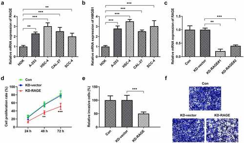
Figure 2. RAGE silencing resulted in the decrease of tube formation in HUVECs. (a) KD-RAGE transfection led to decreased expression of RAGE through RT-qPCR analysis in HSC-4 cells. (b) Effect of RAGE on HUVEC lumen formation ability. ***p < 0.001
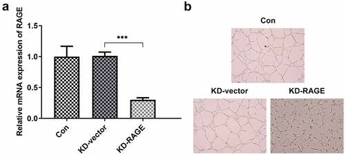
Figure 3. RAGE overexpression abolished the effects of EVO on proliferation and invasion in HSC-4 cells. (a) The proliferation levels of HSC-4 cells were detected by the treatment of 1, 2, 4, 8 or 16 µmol/L. (b) The detection of RAGE mRNA levels, (c) cell proliferation, (d–e) cell invasion when HSC-4 cells were treated with EVO or Ov-RAGE plasmid treatment. ***p < 0.001
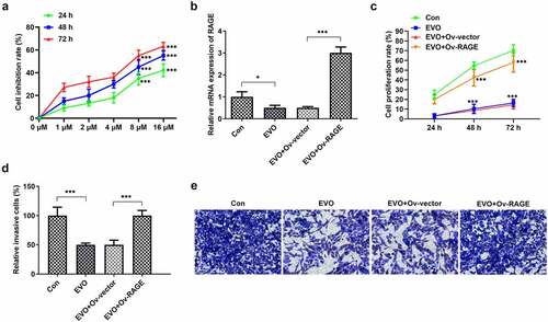
Figure 4. RAGE overexpression blunted the effects of EVO on tube formation in HUVEC cells. (a) Expression of RAGEs was increased when cells were cultured with RAGE overexpressing plasmids. (b) The tube formation abilities of HUVEC cells were analyzed through tuber formation assay. ***p < 0.001
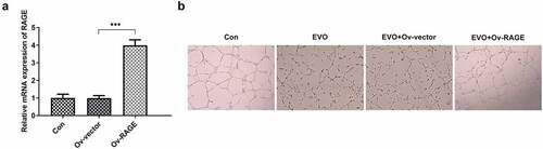
Figure 5. The analysis of HMBG1, RAGE, c-Jun, NF-κB, MMP-2, VEGF and IL-6 expression in vitro after EVO treatment or RAGE overexpression induction. *p < 0.05, **p < 0.01, ***p < 0.001
Figure 6. EVO administration suppressed tumor growth and vasculogenesis via RAGE. (a) The tumor volume, (b) mice weight, (c) tumor weight, (d) photos of mice and (e) Hemotoxylin and eosin staining. *p < 0.05, **p < 0.01, ***p < 0.001
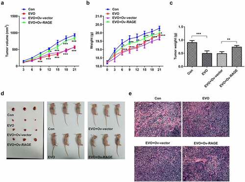
Figure 7. The levels of HMBG1, RAGE, c-Jun, NF-κB, MMP-2, VEGF and IL-6 were affected by EVO treatment, which was mediated by RAGE in vivo. *p < 0.05, **p < 0.01, ***p < 0.001