Figures & data
Table 1. The correlation between LMTK3 expression and clinicopathological features in CRC patients
Figure 1. LMTK3 is highly expressed in CRC. (a) LMTK3 level in colon adenocarcinoma (COAD) and tumor tissues was provided by GEPIA database (P = 0.0342). (b) LMTK3 level in CRC tumors and normal tissues in GSE10950 (P = 0.00647) and GSE74602 (P = 0.00875) datasets from GEO database. (c and d) The mRNA and protein expressions of LMTK3 in 56-paired CRC tissues and adjacent normal tissues was analyzed by RT-qPCR (P = 0.00895) and western blotting (P = 0.00762). (e) LMTK3 mRNA expression in CRC cell lines (LS174T, SW837, HT-29, RKO, and HR8348) and normal colonic epithelial cell line (NCM460) were analyzed by RT-qPCR. (f) ROC curve for prognostic value of LMTK3 in CRC, with AUC = 0.908. *P < 0.05, **P < 0.01
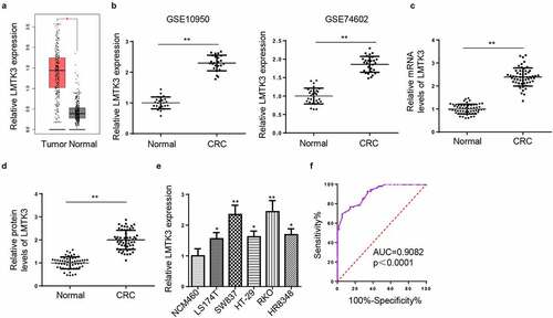
Figure 2. LMTK3 knockdown decreases CTX resistance and cell proliferation and induces cell apoptosis and cell-cycle arrest in CTX-resistant CRC cells. (a) LMTK3 mRNA expression in CTX-sensitive CRC tissues (n = 32) and CTX-resistant CRC tissues (n = 24) was analyzed by RT-qPCR. (b) CTX IC50 values of CTX-resistant CRC cells (RKO/CTX and SW837/CTX) and parental CRC cells (RKO and SW837) were determined by the CCK-8 assay. (c and d) LMTK3 mRNA and protein expressions in CTX-resistant CRC cells (RKO/CTX and SW837/CTX) and parental CRC cells (RKO and SW837) were analyzed by RT-qPCR and western blotting. (e and f) RKO/CTX and SW837/CTX cells were respectively transfected with si-LMTK3 or si-NC, and the transfection efficiency was evaluated by RT-qPCR and western blotting. (g) CTX IC50 values of RKO/CTX and SW837/CTX cells respectively transfected with si-LMTK3 and si-NC were determined by CCK-8 assay. (h) CCK-8 assay was performed to determine cell vitality. (i) Flow cytometry was performed to detect cell apoptosis. (j) Cleaved caspase-3 level was detected by Western blotting. (k) Flow cytometry was performed to detect cell-cycle. *P < 0.05 and **P < 0.01
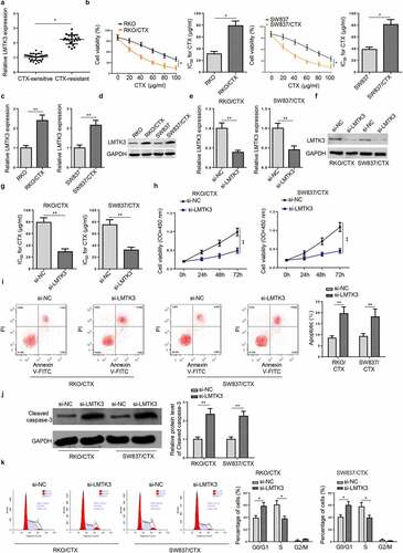
Figure 3. LMTK3 deficiency attenuates CTX-resistant CRC cell migration and invasion. RKO/CTX and SW837/CTX cells were respectively transfected with si-LMTK3 or si-NC. (a) Wound healing was applied to detect cell migration. (b) Transwell assay was applied to assess cell invasion. **P < 0.01 and ***P < 0.001
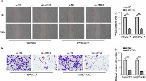
Figure 4. LMTK3 activates ERK/MAPK pathway in CTX-resistant CRC cells. (a) RKO/CTX and SW837/CTX cells were respectively transfected with Vector or oe-LMTK3, and the transfection efficiency was evaluated by western blotting. (b) ERK/MAPK pathway-related proteins (p-ERK1/2 and ERK1/2) in RKO/CTX and SW837/CTX cells transfected with si-LMTK3 or si-NC were analyzed by western blotting. (c) Protein levels of p-ERK1/2 and ERK1/2 in RKO/CTX and SW837/CTX cells transfected with Vector or oe-LMTK3 were analyzed by western blotting. **P < 0.01
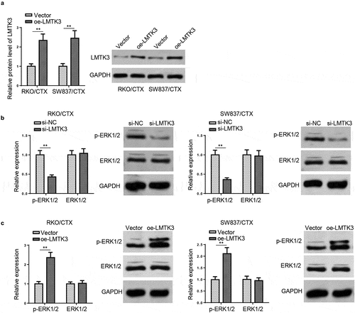
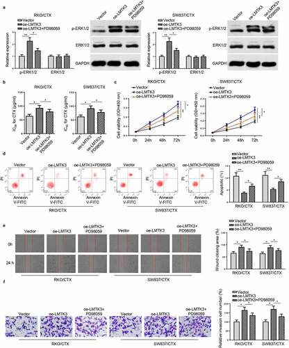
Figure 5. Inactivation of ERK/MAPK signaling reverses the enhanced CTX resistance induced by LMTK3 overexpression in vitro. (a) Protein levels of p-ERK1/2 and ERK1/2 in RKO/CTX and SW837/CTX cells transfected with Vector, oe-LMTK3, or oe-LMTK3+ PD98059 were analyzed by western blotting. (b) CTX IC50 values of RKO/CTX and SW837/CTX cells respectively transfected with Vector, oe-LMTK3, or oe-LMTK3+ PD98059 were determined by CCK-8 assay. (c) CCK-8 assay was performed to determine cell vitality. (d) Flow cytometry was performed to detect cell apoptosis. (e and f) Cell migration and invasion were assessed by wound healing and transwell assays. *P < 0.05 and ** P < 0.01