Figures & data
Figure 1. Ozone prevents cardiomyocytes from injury caused by H/R in vitro. (a) Treat various concentrations (0, 5, 10, 20, 30, 40, 60, 80 μg/ml) of ozone to the cardiomyocytes of primary rat and choose CCK-8 analysis to test cell viability. (b) Treat with 10 μg/ml or 20 μg/ml to cardiomyocytes after 4 hours of hypoxia, and then incubate in a normal incubator with oxygen for 6 hours to reoxygenate, and measure the viability of cell by CCK-8. (c) Release level of LDH. (d) CK activity. (e) analysis of Western blot about the levels of protein of Bax, cleaved caspase-3, Bcl-2 and caspase-3. (f) Quantification of Bax/Bcl-2 ratio. (g) Activity level of caspase-3. n = 3 *p < 0.05, **p < 0.01
Figure 2. In animal models, ozone attenuated myocardial injury caused by I/R. (a) The LDH level of Serum. (b) CK-MB activity of Serum. (c) Representative photographs in TTC staining for heart tissues and the myocardial infarct size was calculated. (d) Levels of protein of Bax, caspase-3, cleaved caspase-3 and Bcl-2 were detected by Western blot. (e) Quantification of Bax/Bcl-2 ratio. (f) Activity level of caspase-3. n = 3 **p < 0.01
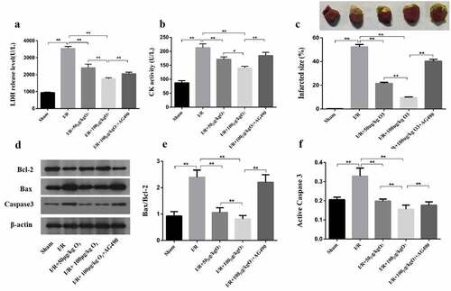
Figure 3. Ozone inhibited the injury of cardiomyocyte caused by H/R through activating the signaling of JAK2/STAT3. (a) Western blot to detect the protein levels of STAT3, p-JAK2, p-STAT3 and JAK2. (b) Quantification of p-STAT3/STAT3 ratios. (c) Quantification of p-JAK2/JAK2 ratios. 30 min before applying the H/R, cardiomyocytes, treated with JAK2 inhibitor AG-490 (2 mM), were incubated during the whole process of H/R. (d) The release level of LDH. (e) CK activity. (f) After assaying, the levels of expression of Bax, caspase-3, cleaved caspase-3 and Bcl-2 were determined by Western blot. (g) Quantification of Bax/Bcl-2 ratio. (h) Activity level of caspase-3. each group n = 3, **p < 0.01
Figure 4. The effect of ozone mediated by HSP70 to Cardiomyocytes Injury induced by I/R. (a) The relative expression of HSP70 mRNA was detected by RT-PCR. (b) The protein level of HSP70 was assayed by Western blot. (c) Cardiomyocytes were transfected by HSP70 siRNA, control or pretreated with AG-490 followed by 20 μg/ml ozone treatment and I/R treatment. HSP70 protein level was determined. LDH release level (d) and CK activity (e) in Cardiomyocytes were detected. n = 3 per group, **p < 0.01
Figure 5. Ozone alleviated myocardial injury caused by I/R through up-regulating HSP70 and activating JAK2/STAT3 in vivo. (a) Analyzed the levels of protein of JAK2, p-STAT3, p-JAK2, HSP70 and STAT3 in I/R animal models. (b–d) Quantification of HSP70 level, as well as p-STAT3/STAT3 and p-JAK2/JAK2 ratios. n = 6, **p < 0.01