Figures & data
Table 1. Clinicopathologic details of the patients
Figure 1. High APCDD1L-AS1 is discovered in OSCC and correlative to poor prognosis of OSCC patients
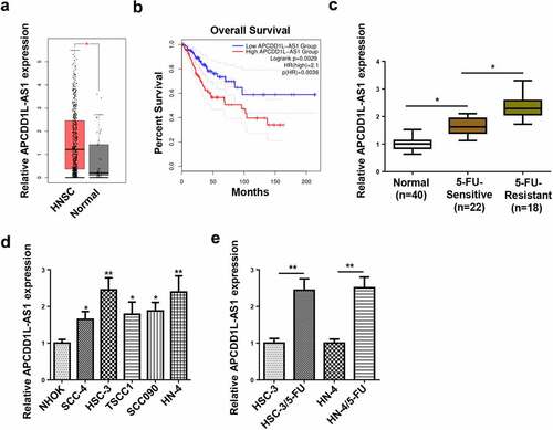
Figure 2. APCDD1L-AS1 confers resistance to 5-FU in OSCC cells
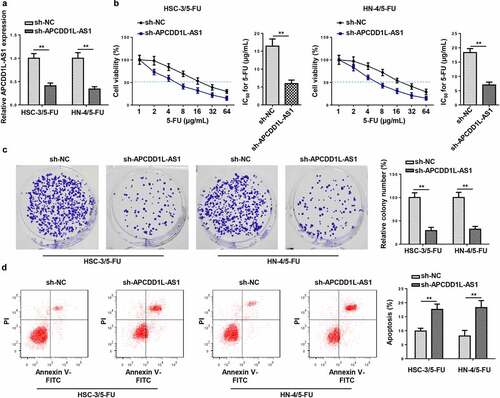
Figure 3. APCDD1L-AS1 is transferred extracellularly via exosome incorporation in 5-FU-Resistant OSCC Cells
Figure 4. APCDD1L-AS1 inhibition attenuates 5-FU resistance mediated by exosomes
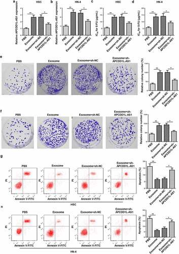
Figure 5. APCDD1L-AS1 serves as a sponge for miR-1224-5p in 5-FU-resistant OSCC cells
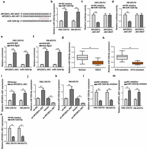
Figure 6. MiR-1224-5p overexpression overcomes 5-FU resistance in 5-FU-resistant OSCC cells
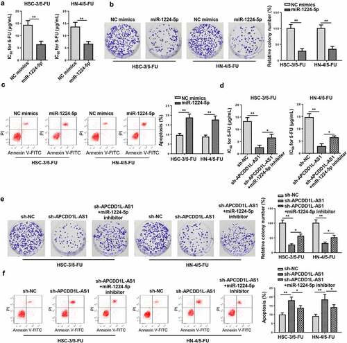
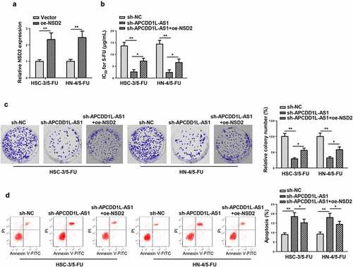
Figure 7. NSD2 is a direct target of miR-1224-5p in 5-FU-resistant OSCC cells
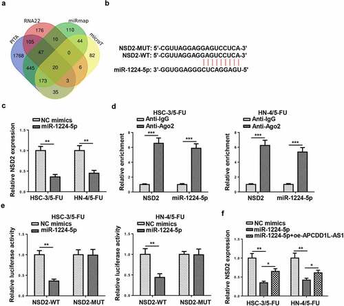
Figure 8. APCDD1L-AS1 knockdown enhances susceptibility to 5-FU in 5-FU-resistant OSCC cells by decreasing NSD2