Figures & data
Figure 1. Expressions of CCNG1/TP53/MMP7 and LncRNA CASC9/miR-488-3p were abnormal in OC cells. A, Protein levels of CCNG1, TP53 and MMP7 in several OC cells were detected by western blot analysis. B-C, mRNA levels of CASC9 and miR-488-3p in several OC cells were measured by qRT-PCR. Data are expressed as mean ± SD. *P < 0.05, ***P < 0.001 versus IOSE-80 cells
Figure 2. CASC9 promoted cell proliferation, migration and invasion in OVCAR-3 and OVCAR-3/DDP cells. A, Cell inhibitory rate in OVCAR-3 and OVCAR-3/DDP cells was evaluated by CCK-8 assay. *P < 0.05. B, CASC9 mRNA level was detected after transfection with shRNA-CASC9 or OverExp-CASC9. ***P < 0.001 versus control; ###P < 0.001 versus vector. C, Cell proliferation were identified by CCK-8 assay after transfection with shRNA-CASC9 or OverExp-CASC9. D, Cell inhibitory rate was identified by CCK-8 assay after transfection with shRNA-CASC9 or OverExp-CASC9. E, Cell migration was evaluated by wound healing assay after transfection with shRNA-CASC9 or OverExp-CASC9. F, Cell invasion was examined by transwell assay after transfection with shRNA-CASC9 or OverExp-CASC9. Data are expressed as mean ± SD. *P < 0.05, ***P < 0.001 versus OVCAR-3 cells; #P < 0.05, ##P < 0.01, ###P < 0.001 versus OVCAR-3/DDP+vector
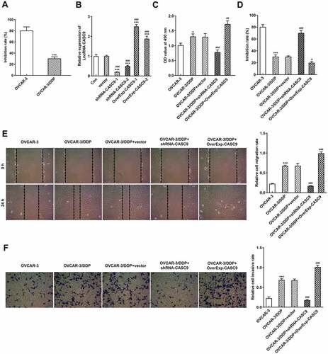
Figure 3. CASC9 suppressed cell apoptosis and cell cycle in OVCAR-3 and OVCAR-3/DDP cells. A, Tunel assay was performed to assess cell apoptosis after transfection with shRNA-CASC9 or OverExp-CASC9. B, Protein levels of Bax, Bcl-2, caspase 3 and cleaved caspase 3 were measured by western blot analysis after transfection with shRNA-CASC9 or OverExp-CASC9. C, Flow cytometric analysis was carried out to detect cell apoptosis after transfection with shRNA-CASC9 or OverExp-CASC9. Data are expressed as mean ± SD. **P < 0.01, ***P < 0.001 versus OVCAR-3 cells; #P < 0.05, ###P < 0.001 versus OVCAR-3/DDP+vector
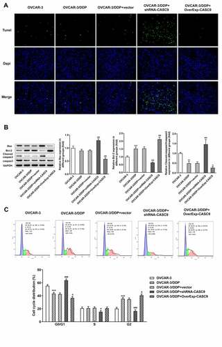
Figure 4. Expressions of CCNG1/TP53/MMP7 were suppressed by CASC9 silencing. Western blot analysis was performed to identify the protein levels of CCNG1, TP53 and MMP7 in OVCAR-3 and OVCAR-3/DDP cells transfected with shRNA-CASC9 or OverExp-CASC9. Data are expressed as mean ± SD. ***P < 0.001 versus OVCAR-3 cells; #P < 0.05, ##P < 0.01, ###P < 0.001 versus OVCAR-3/DDP+vector
Figure 5. CASC9/miR-488-3p directly targets CCNG1. A-B, mRNA levels of CASC9 and miR-488-3p were identified by qRT-PCR after transfection with shRNA-CASC9 or OverExp-CASC9. C, The binding sequence of CASC9 and miR-488-3p. D, Luciferase reporter assay was performed to verify the combination between CASC9 and miR-488-3p. E, The binding sequence of miR-488-3p and CCNG1. F, Luciferase reporter assay was performed to verify the combination between miR-488-3p and CCNG1. Data are expressed as mean ± SD. *P < 0.05, ***P < 0.001 versus control; ##P < 0.01, ###P < 0.001 versus OVCAR-3/DDP+vector
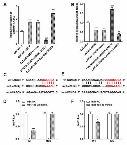
Figure 6. Effects of miR-488-3p overexpression on cell resistance and apoptosis in OVCAR-3/DDP cells. A, Cell inhibitory rate was detected by CCK-8 assay after transfection with OverExp-CASC9 in the presence and absence of miR-488-3p mimic. B, Tunel assay was carried out to assess cell apoptosis after transfection with OverExp-CASC9 in the presence and absence of miR-488-3p mimic. Data are expressed as mean ± SD. ***P < 0.01 versus vector; ##P < 0.01, ###P < 0.001 versus OverExp-CASC9+ mimic NC
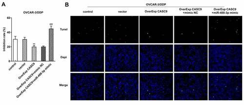
Figure 7. Effects of CASC9 silencing or overexpression on tumor growth and apoptosis in vivo. A, Ovarian cancer xenograft mouse model was established. B, Tumor weight of mice model was measured after transfection with shRNA-CASC9 or OverExp-CASC9. C, Tumor volume of mice model was evaluated after transfection with shRNA-CASC9 or OverExp-CASC9. D, Cell apoptosis was assessed in tumor tissues of mice by tunel assay after transfection with shRNA-CASC9 or OverExp-CASC9. Data are expressed as mean ± SD. ***P < 0.001 versus OVCAR-3 cells; ###P < 0.001 versus OVCAR-3/DDP+vector
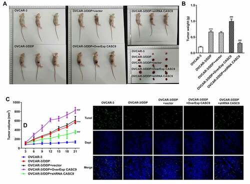
Figure 8. Effects of CASC9 on protein expressions involved in CCNG1/TP53/MMP7 and apoptosis. A, Protein levels of CCNG1, TP53 and MMP7 in tumor tissues of mice was evaluated by western blot assay transfected with shRNA-CASC9 or OverExp-CASC9. B, Protein levels of Bax, Bcl-2, caspase 3 and cleaved caspase 3 in tumor tissues of mice were identified by western blot analysis after transfection with shRNA-CASC9 or OverExp-CASC9. C-D, Expression of CASC9 and miR-488-3p in tumor tissues of mice was determined with qRT-PCR after transfection with shRNA-CASC9 or OverExp-CASC9. Data are expressed as mean ± SD. *p < 0.05, **p < 0.01, ***P < 0.001 versus OVCAR-3 cells; #P < 0.05, ##P < 0.01, ###P < 0.001 versus OVCAR-3/DDP+vector