
Figures & data
Table 1. Significance of blood-derived EV-related molecules in diseases
Figure 1. The function of erythrocyte-derived extracellular vesicles (REVs) REVs elicit immune-inflammatory responses by modulating the biological activities of both T cells and B cells. REVs stimulate monocytes to produce proinflammatory cytokines and chemokines, which promote T cell proliferation and further stimulate T cells to produce TNF, IL-6, and IL-8. REVs also inhibit the expression of Blimp-1 and IRF4 and activation of the NF-κB pathway, which inhibit B cell function. Additionally, REVs mediate blood coagulation by activating coagulation factors such as FXI, which initiates and promotes thrombin production
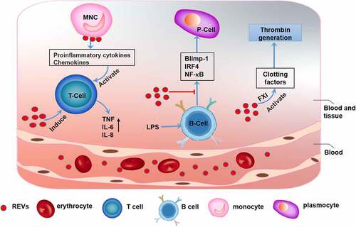
Table 2. Summary of blood cell-derived EVs for drug delivery
Figure 2. Characterization of erythrocyte-derived extracellular vesicles (REVs)

Figure 3. The function of leukocyte-derived extracellular vesicles (LEVs)
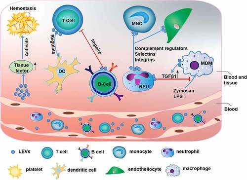
Figure 4. The function of platelet-derived extracellular vesicles (PEVs)
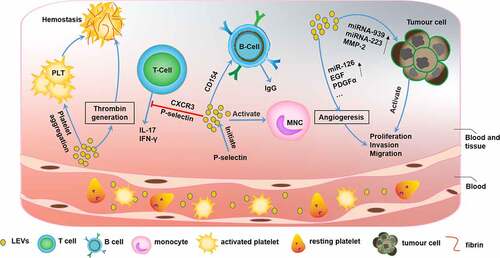
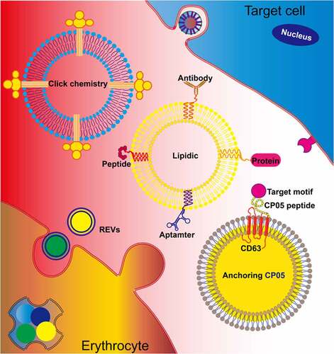
Figure 5. Erythrocyte-derived extracellular vesicles (REVs) for therapeutic delivery To optimize the targeted delivery of REVs, chemical modifications introduced by approaches such as click chemistry and lipidation can anchor the targeted motifs, including proteins, aptamers, peptides, and antibodies, on the exosomal membrane. Additionally, REVs can be modified using the CP05 peptide, with affinity for CD63, to introduce an exogenous target motif for display on the exosomal membrane
Data Availability Statement
All data generated in the current study were included in this article.