
Figures & data
Figure 1. The expression of ANGPT2 was increased in IEC-6 cells after LPS induction. (a) IEC cells were incubated with different concentrations of LPS (0, 0.1, 1, 10, and 100 μg/ml) for 24 h. The cell viability of LPS-induced IEC-6 cells was detected using CCK-8. (b) The mRNA level of ANGPT2 in LPS-induced IEC-6 cells was detected using RT-qPCR. (c) The protein expression of ANGPT2 in LPS-induced IEC-6 cells was detected using western blot. *p < 0.05 and ***p < 0.001 vs Control
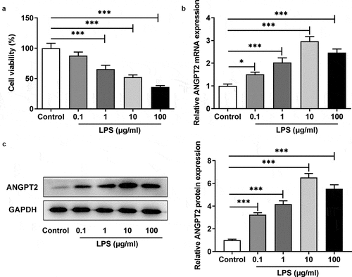
Figure 2. ANGPT2 knockdown inhibited the apoptosis of LPS-induced IEC-6 cells. (a) IEC-6 cells were transfected with sh-NC or sh-ANGPT2-1/2, and the mRNA expression and (b) the protein expression of ANGPT2 in IEC-6 cells were detected by RT-qPCR and western blot, respectively. (c) The transfected or untransfected IEC-6 cells were induced with LPS (10 μg/ml) for 24 h. The cell viability of was detected using CCK-8. (d) The cell apoptosis of each group was checked by TUNEL. ***p < 0.001
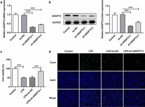
Figure 3. ANGPT2 knockdown inhibited the inflammatory response, barrier dysfunction and ER stress of LPS-induced IEC-6 cells. (a–d) The transfected or untransfected IEC-6 cells were induced with LPS (10 μg/ml) for 24 h. The levels of PGE2, TNF-α, IL-1β and IL-6 were checked by ELISA. (e) The cell monolayer permeability was detected using TEER. (f) The expressions of ZO-1, occludin and claudin-1 were measured by western blot. (g) The expressions of GRP79, CHOP, p-PERK and PERK were measured by western blot. *p < 0.05, **p < 0.01, ***p < 0.001
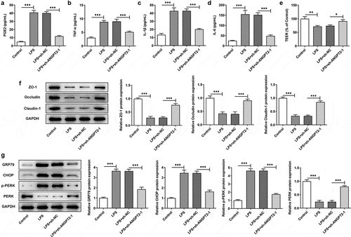
Figure 4. ANGPT2 knockdown blocked Notch signaling pathway. The transfected or untransfected IEC-6 cells were induced with LPS (10 μg/ml) for 24 h. The expressions of NICD1 and HES1 were measured using western blot. ***p < 0.001
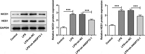
Figure 5. ANGPT2 knockdown inhibited the apoptosis of LPS-induced IEC-6 cells through suppressing Notch signaling pathway. (a) The transfected or untransfected IEC-6 cells were induced with LPS (10 μg/ml) for 24 h. Jagged-1 (JAG), an agonist of Notch, was used for treatment. The cell viability was detected using CCK-8. (b) The cell apoptosis of each group was checked using TUNEL. **p < 0.01, ***p < 0.001
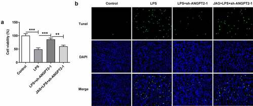
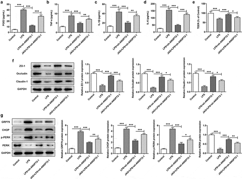
Figure 6. ANGPT2 knockdown improved the inflammatory response, barrier dysfunction and ER stress of LPS-induced IEC-6 cells via suppressing Notch signaling pathway. (a–d) The transfected or untransfected IEC-6 cells were induced with LPS (10 μg/ml) for 24 h. Jagged-1 (JAG), an agonist of Notch, was used for treatment. The levels of PGE2, TNF-α, IL-1β and IL-6 were checked by ELISA. (e) The cell monolayer permeability was detected using TEER. (f) The expressions of ZO-1, occludin and claudin-1 were measured by western blot. (g) The expressions of GRP79, CHOP, p-PERK and PERK were measured by western blot. *p < 0.05, **p < 0.01, ***p < 0.001
Data availability statement
All data in this study have been included in this article.