
Figures & data
Table 1. The relationship between AURKA expression and clinical characteristics
Table 2. Primer for qRT-PCR
Figure 1. AURKA presented a high expression in DLBCL tissues and cells. (a) The expression of AURKA in the DLBC tumor tissues from DLBC patients than that in normal lymphoid tissues from healthy people was analyzed by GEPIA2 (http://gepia2.cancer-pku.cn/#index). (b) The expression of AURKA in DLBCL tumor tissues from DLBCL patients and normal lymphoid tissues from healthy volunteers was detected using qRT-PCR, and the GAPDH was the internal control. (c) The expression of AURKA in DLBCL tumor tissues from DLBCL patients and normal lymphoid tissues from healthy volunteers was detected using immunohistochemistry (IHC) assay (magnification, ×100). (d) The expression of AURKA in DLBCL cells and normal B lymphocyte was detected using qRT-PCR, and the GAPDH was the internal control. All experiments were repeatedly performed over 3 times. Experimental data were expressed by mean ± standard deviation (SD). (*P < 0.05, ***P < 0.001; ^^^P < 0.001; * vs. normal group; ^ vs. GM12878 group) (AURKA: Aurora-kinase-A; DLBCL: diffuse large B-cell lymphoma; GEPIA2:gene expression profiling interactive analysis 2; qRT-PCR: quantitative real-time polymerase chain reaction)
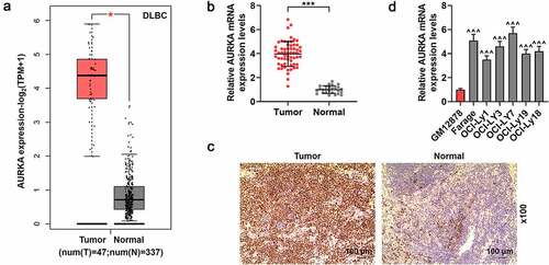
Figure 2. Silencing AURKA negatively regulated AURKA expression and viability of DLBCL cells, and promoted the apoptosis of DLBCL cells. (a) The mRNA expression of AURKA in OCI-LY7 cells transfected with siAURKA or siNC was detected by qRT-PCR, and the GAPDH was internal control. (b-c) The protein expression of AURKA in OCI-LY7 cells transfected with siAURKA or siNC was detected by Western blot, with GAPDH as the internal control. (d) The mRNA expression of AURKA in Farage cells transfected with siAURKA or siNC was detected by qRT-PCR, and the GAPDH was used as the internal control. (e-f) The protein expression of AURKA in Farage cells transfected with siAURKA or siNC was detected by Western blot, and the GAPDH was the internal control. (g-h) The cell viability of OCI-LY7 (g) and Farage (h) cells transfected with siAURKA or siNC and cultured for 24 h and 48 h was detected by CCK-8 assay. (i-l) The cell apoptosis of OCI-LY7 (i-j) and Farage (k-l) cells transfected with siAURKA or siNC was detected by flow cytometry. All experiments were repeatedly performed over 3 times. Experimental data were expressed by mean ± standard deviation (SD). (*P < 0.05, **P < 0.01, ***P < 0.001; * vs. siNC group) (AURKA: Aurora-kinase-A; DLBCL: diffuse large B-cell lymphoma; siAURKA: short interfering RNA targeting AURKA; siNC: negative control of siAURKA; qRT-PCR: quantitative real-time polymerase chain reaction; CCK-8: cell counting kit 8; h: hours)
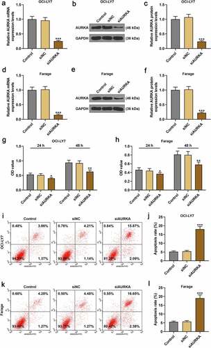
Figure 3. CHOP reduced the viability and promoted apoptosis of DLBCL cells, and silencing AURKA enhanced the effects of CHOP on DLBCL cell apoptosis. (a-b) After treatment with CHOP (0, 10, 20 and 40 ng/mL) and cultured for 24 h and 48 h, the cell viability of OCI-LY7 (a) and Farage (b) cells was detected by CCK-8 assay. (c-d) After treatment with CHOP (0, 10, 20 and 40 ng/mL), the expression of AURKA in OCI-LY7 (c) and Farage (d) cells was detected by qRT-PCR, and GAPDH worked as internal control. (e-f) After transfection with siAURKA or siNC and treatment with CHOP (20 ng/mL), the apoptosis of OCI-LY7 (e) and Farage (f) cells was detected by flow cytometry. All experiments were repeatedly performed over 3 times. Experimental data were expressed by mean ± standard deviation (SD). (**P < 0.01, ***P < 0.001; ^^^P < 0.001; ###P < 0.001; &&&P < 0.001; * vs. Control group; ^ vs. siNC group; # vs. siAURKA group; & vs. siNC+CHOP group) (CHOP: cyclophosphamide, doxorubicin, vincristine, and prednisone; AURKA: Aurora-kinase-A; DLBCL: diffuse large B-cell lymphoma; qRT-PCR: quantitative real-time polymerase chain reaction; siAURKA: short interfering RNA targeting AURKA; siNC: negative control of siAURKA; CCK-8: cell counting kit 8; h: hours)
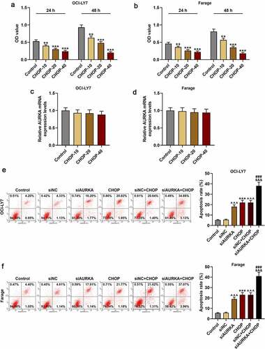
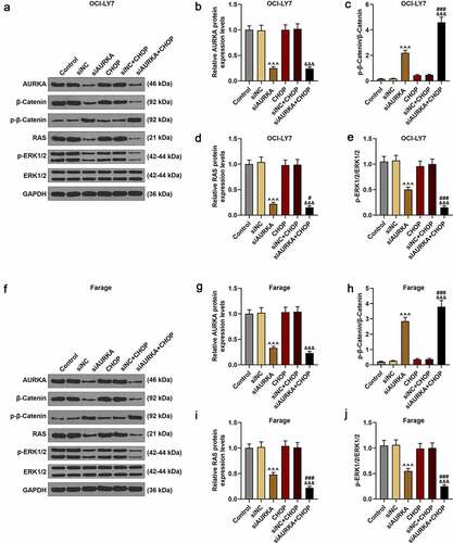
Figure 4. Silencing AURKA downregulated the AURKA and RAS expressions, reduced the ratio of p-ERK1/2/ERK1/2 yet increased the ratio of p-β-Catenin/β-Catenin, and these effects were reinforced by CHOP. (a-j) After the OCI-LY7 (a-e) and Farage (f-j) cells were transfected with siAURKA or siNC and treated with CHOP (20 ng/mL), the protein expressions of AUKA, RAS, p-β-Catenin, β-Catenin, p-ERK1/2 and ERK1/2 were detected using Western blot and the ratios of p-β-Catenin/β-Catenin and p-ERK1/2/ERK1/2 were analyzed, with GAPDH used as an internal control. All experiments were repeatedly performed over 3 times. Experimental data were expressed by mean ± standard deviation (SD). (^^^P < 0.001; #P < 0.05, ###P < 0.001; &&&P < 0.001; ^ vs. siNC group; # vs. siAURKA group; & vs. siNC+CHOP group) (CHOP: cyclophosphamide, doxorubicin, vincristine, and prednisone; AURKA: Aurora-kinase-A; DLBCL: diffuse large B-cell lymphoma; siAURKA: short interfering RNA targeting AURKA; siNC: negative control of siAURKA; ERK: extracellular-signal regulated kinase; p-ERK: phosphorylated-ERK)
Availability of data and materials
The analyzed data sets generated during the study are available from the corresponding authors on reasonable requests.