
Figures & data
Table 1. Clinicopathological features of subjects
Figure 1. MiR–143–3p is lifted in RPL and impairs the biological functions of trophoblastic cells. (a) Relative miR–143–3p expression in placental tissues from RPL patients (n = 28) and normal pregnant women (n = 28) was detected by RT-qPCR. (b) miR–143–3p expression in HTR8/SVneo cells transfected with NC mimics, NC inhibitor, or miR–143–3p inhibitor was detected by RT-qPCR. (c) The cell viability of transfected HTR8/SVneo cells was analyzed by CCK-8 assay at different time points. (d) The apoptosis condition of transfected HTR8/SVneo cells was analyzed by flow cytometry assay. (e) The migration capability of transfected HTR8/SVneo cells was analyzed by wound healing assay. (f) The invasion capability of transfected HTR8/SVneo cells was analyzed by transwell assay. (g and h) The mRNA or protein expression level of EMT markers (E-cadherin, vimentin, and N-cadherin) in transfected HTR8/SVneo cells was determined by RT-qPCR or western blotting. Data are shown as the mean ± SD (n = 3). *P < 0.05; **P < 0.01
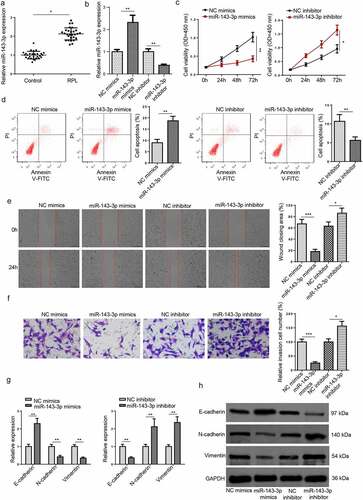
Figure 2. S100A11 is directly targeted by miR–143–3p in trophoblastic cells. (a) 8 candidate target mRNAs for miR–143–3p were predicted by using three miRNA databases (miRanda, miRmap, and PITA). (b) Relative S100A11 expression in placental tissues from RPL patients (n = 28) and normal pregnant women (n = 28) was detected by RT-qPCR. (c) The putative binding sites between miR–143–3p and S100A11. (d) The HTR8/SVneo cells were co-transfected with either S100A11-WT or S100A11-MUT, and miR–143–3p mimics/inhibitor or corresponding NC. Then, the relative luciferase activity was measured. (e) Pearson’s correlation analysis indicated a negative correlation between the expression of miR–143–3p and S100A11 in placental tissues from RPL patients (n = 28). (f and g) S100A11 mRNA or protein level in HTR8/SVneo cells transfected with NC mimics, miR–143–3p mimics, NC inhibitor or miR–143–3p inhibitor was detected by RT-qPCR or western blotting. Data are shown as the mean ± SD (n = 3). *P < 0.05; **P < 0.01
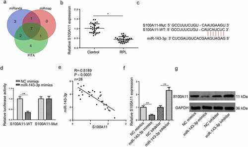
Figure 3. MiR–143–3p weakens trophoblastic cell functions via S100A11. (a and b) S100A11 mRNA or protein level in HTR8/SVneo cells transfected with si-NC or si-S100A11 was detected by RT-qPCR or western blotting. (c) HTR8/SVneo cells transfected with NC inhibitor, miR–143–3p inhibitor, miR–143–3p inhibitor+si-NC, or miR–143–3p inhibitor+si-S100A11, respectively. the cell viability of transfected HTR8/SVneo cells was analyzed by CCK-8 assay at different time points. (d) The apoptosis condition of transfected HTR8/SVneo cells was analyzed by flow cytometry assay. (e) The migration capability of transfected HTR8/SVneo cells was analyzed by wound healing assay. (f) The invasion capability of transfected HTR8/SVneo cells was analyzed by transwell assay. (g and h) The mRNA or protein expression level of S100A11 and EMT markers (E-cadherin, vimentin, and N-cadherin) in transfected HTR8/SVneo cells were determined by RT-qPCR or western blotting. data are shown as the mean ± SD (n = 3). *P < 0.05; **P < 0.01
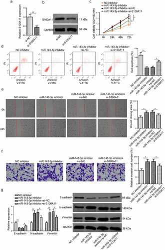
Figure 4. CircFOXP1 binds to miR–143–3p in trophoblastic cells. (a) The putative binding sites between miR–143–3p and circFOXP1. (b) The HTR8/SVneo cells were co-transfected with either circFOXP1-WT or circFOXP1-MUT, and miR–143–3p mimics or NC mimics. then, the relative luciferase activity was measured. (c) Relative S100A11 expression in placental tissues from RPL patients (n = 28) and normal pregnant women (n = 28) was detected by RT-qPCR. (d) CircFOXP1 expression in HTR8/SVneo cells transfected with si-NC or si-circFOXP1 was detected by RT-qPCR. (e) CircFOXP1 expression in HTR8/SVneo cells transfected with vector or oe-circFOXP1was detected by RT-qPCR. (f) Pearson’s correlation analysis indicated a negative correlation between the expression of miR–143–3p and circFOXP1 in placental tissues from RPL patients (n = 28). (g) miR–143–3p expression in HTR8/SVneo cells transfected with si-NC, si-circFOXP1, Vector, or oe-circFOXP1 was detected by RT-qPCR. (h) Pearson’s correlation analysis indicated a positive correlation between S100A11 mRNA expression and circFOXP1 expression in placental tissues from RPL patients (n = 28). (i and j) S100A11 mRNA or protein level in HTR8/SVneo cells transfected with si-NC, si-circFOXP1, Vector, or oe-circFOXP1 was detected by RT-qPCR or western blotting. data are shown as the mean ± SD (n = 3). *P < 0.05; **P < 0.01
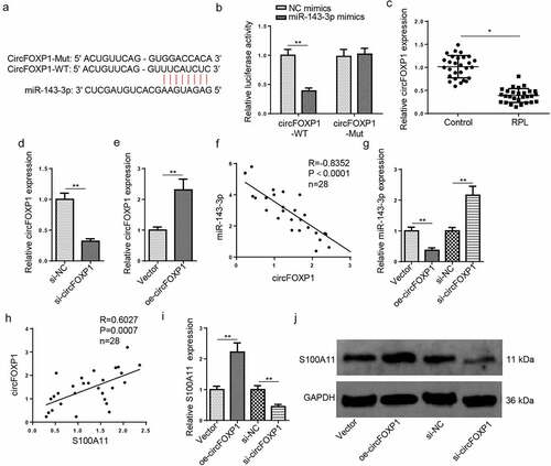
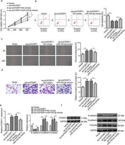
Figure 5. CircFOXP1/miR–143–3p/S100A11 axis regulates trophoblastic cell functions. (a) HTR8/SVneo cells transfected with vector, oe-circFOXP1, oe-circFOXP1+NC mimics, or oe-circFOXP1+ miR–143–3p mimics, respectively. The cell viability of transfected HTR8/SVneo cells was analyzed by CCK-8 assay at different time points. (b) The apoptosis condition of transfected HTR8/SVneo cells was analyzed by flow cytometry assay. (c) The migration capability of transfected HTR8/SVneo cells was analyzed by wound healing assay. (d) The invasion capability of transfected HTR8/SVneo cells was analyzed by transwell assay. (e and f) The mRNA or protein expression level of S100A11 and EMT markers (E-cadherin, vimentin, and N-cadherin) in transfected HTR8/SVneo cells were determined by RT-qPCR or western blotting. data are shown as the mean ± SD (n = 3). *P < 0.05; **P < 0.01
Data availability statement
The datasets used and/or analyzed during the current study are available from the corresponding author on reasonable request.