Figures & data
Figure 1. PTP1B expression is highly elevated in malignant melanoma and correlates with poor survival
A. PTP1B mRNA expression analysis in MM, nevi and normal tissues (GSE3189; Normal, n = 7; Nevi, n = 18; Melanoma, n = 45).B. PTP1B mRNA expression analysis in primary and metastatic tissues (GSE8401; Primary, n = 31; Metastasis, n = 52).C. PTP1B mRNA expression analysis in normal, primary and metastatic tissues (GSE15605; Normal, n = 16; Primary, n = 46; Metastasis, n = 12)D. Overall survival of MM patients based on PTP1B mRNA expression (GSE19234; High, n = 23; Low, n = 21).E. PTP1B protein expression in nontumor and melanoma tissues by immunohistochemical AEC staining. Original magnification 400 × .F. PTP1B expression was analyzed in nontumor and melanoma tissues (Normal, n = 18; Melanoma, n = 82).
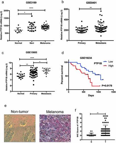
Figure 2. PTP1B deficiency inhibits the horizontal migration of melanoma cells without affecting cell proliferation
A The proliferation ability of A2058 and MV3 cells with knockdown of PTP1B was assessed by the CCK-8 assay (OD value 450 nm).B The proliferation ability of A2058 and MV3 cells with overexpression of PTP1B was assessed by the CCK-8 assay (OD value 450 nm).C The horizontal migration ability of A2058 cells with knockdown of PTP1B was assessed by the wound healing assay at 48 h.D The horizontal migration ability of A2058cells with overexpression of PTP1B was assessed by the wound healing assay at 48 h.
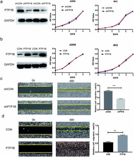
Figure 3. PTP1B-overexpressing melanoma cells show increased vertical migration and invasion
A&B The vertical migration ability of MV3 cells with knockdown or overexpression of PTP1B was assessed by the Transwell migration assay.C&D The invasion ability of A2058 cells with knockdown or overexpression of PTP1B was assessed by the Transwell invasion assay.
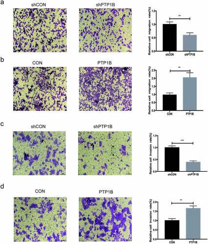
Figure 4. PTP1B promotes tumor metastasis in vivo
A. PET-CT images showing the lung metastasis area (red arrow).B. Analysis of the SUV value in the lung metastases.C. Images of lung metastases subjected to HE staining. Original magnification, 40× and 200 × .D. Analysis of the relative lung metastasis based on the number and area of metastases.
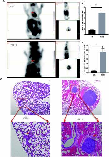
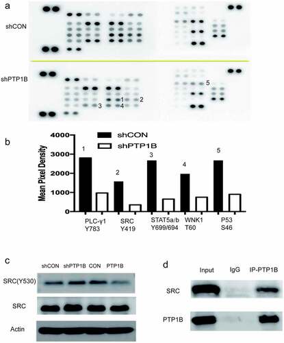
Figure 5. PTP1B regulates melanoma cell migration by interacting with Src
A. Phosphorylation levels of proteins in A2058 cells with knockdown of PTP1B were detected in the Human Phospho-Kinase Array.B. Mean pixel density shows changes in the phosphorylation status of proteins.C. The phosphorylation level of Src Tyr530 was detected in cells with knockdown or overexpression of PTP1B.D. Proteins pulled down with PTP1B-HA antibody were evaluated by western blotting for the presence of PTP1B and Src.