Figures & data
Figure 1. miR-19a-3p and HIF-1α expression. (a) The levels of HIF-1α were determined by WB (b) and qRT-PCR (c) MiR-19a-3p mRNA level was detected through qRT-PCR. (d) Results pointed out miR-19a-3p and HIF-1α levels in KG-1 cells were higher than HS-5 cell lines
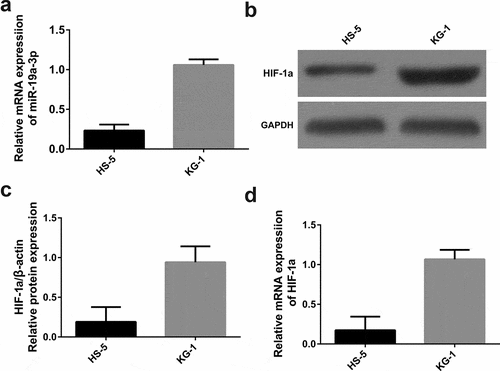
Figure 2. Effects of different concentrations of simvastatin on miR-19a-3p/HIF-1α in KG-1. Different concentrations of simvastatin were added to intervene KG-1 for 24 hours. (a–b) HIF-1α and miR-19a-3p mRNA levels were determined through qRT-PCR. (c–d) HIF-1α protein level was identified by WB
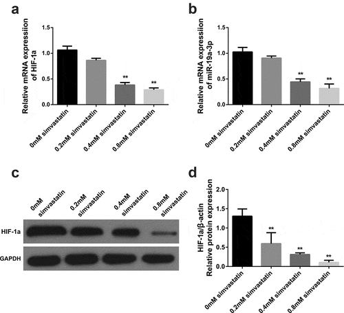
Figure 3. Simvastatin induces miR-19a-3p expression in AML cells to regulate the proliferation, apoptosis, migration and invasion. (a) qRT-PCR served to identify miR‐19a‐3p mRNA of AML cell lines. (b) Cell viability was analyzed by CCK-8. (c) Apoptosis analysis was identified through Flow Cytometry. (d) Apoptosis protein levels were performed by WB. (e) The migration and invasion were observed by transwell and wound healing. Compared with control group, *P < 0.05, **P < 0.01. Compared with the simvastatin group, #P < 0.05, ##P < 0.01
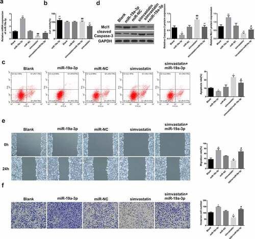
Figure 4. miR-19a-3p directly targets HIF-1α. (a) Luciferase activity was detected by luciferase gene reporting experiment. (b) qPCR severed to identify HIF-1α mRNA levels. (c–d) HIF-1α protein level was performed through WB. Statistical analysis: compared with miR-19a-3p group, *P < 0.05, **P < 0.01. Compared with the miR-19a-3p inhibitor group, #P < 0.05, ##P < 0.01
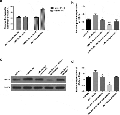
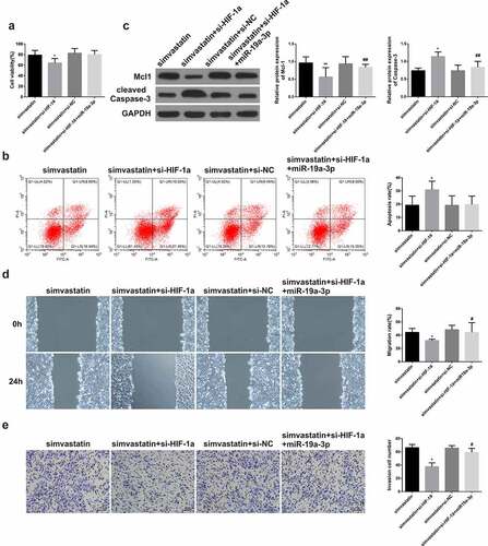
Figure 5. Simvastatin regulates the proliferation, apoptosis, migration and invasion of human acute myeloid leukemia cells via blocking HIF-1α by miR-19a-3p. (a) Cell proliferation was detected through CCK-8. (b) Cell apoptosis analysis was observed via Flow Cytometry. (c) The levels of apoptosis protein of Mcl-1, caspase-3 were performed by WB. (d-e) The migration and invasion were detected by wound healing assay and transwell separately. Statistical analysis: compared with simvastatin group, *P < 0.05, **P < 0.01. Compared with the simvastatin+si-HIF-1α group, #P < 0.05, ##P < 0.01