
Figures & data
Figure 1. NMDA induces autophagy and injury of RGCs. (a) The viability of RGCs treated with different concentrations (0, 50, 100 or 150 μmol/L) of NMDA was evaluated by MTT assay. (b) The expression of autophagy-related proteins (LC3-I, LC3-II, Beclin-1 and ATG5) in RGCs treated with different concentrations (0, 50, 100 or 150 μmol/L) of NMDA was examined by Western blotting. (c) The expression of PI3K/AKT signaling-associated proteins (pAKT, AKT, pPI3K and PI3K) in RGCs treated with different concentrations (0, 50, 100 or 150 μmol/L) of NMDA was tested by Western blotting. *p < 0.05, ** p < 0.01, ***p < 0.001
Figure 2. Baicalin inhibits autophagy and injury of NMDA-treated RGCs via activating PI3K/AKT signaling. (a) The viability of RGCs in four groups (Ctrl, NMDA, NMDA+BAI and NMDA+BAI+LY294002) was assessed by MTT assay. (b-c) Apoptosis of RGCs in four groups (Ctrl, NMDA, NMDA+BAI and NMDA+BAI+LY294002) was tested by flow cytometry. (d-e) The expression of autophagy-related proteins (LC3-I, LC3-II, Beclin-1 and ATG5) and PI3K/AKT signaling-associated proteins (pAKT, AKT, pPI3K and PI3K) in RGCs in four groups (Ctrl, NMDA, NMDA+BAI and NMDA+BAI+LY294002) was evaluated by Western blotting. (f) Chemical structure of baicalin was presented. ** p < 0.01, *** p < 0.001
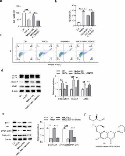
Figure 3. Baicalin restrains NMDA-induced oxidative stress injury of RGCs via activating PI3K/AKT signaling. (a) DCFH-DA staining assay was performed and (b) ROS and (c) MDA levels in RGCs in four groups (Ctrl, NMDA, NMDA+BAI and NMDA+BAI+LY294002) were measured. *p < 0.05, ** p < 0.01
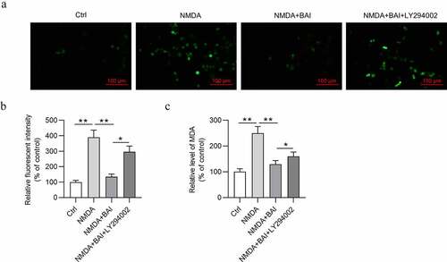
Figure 4. Successful establishment of a mouse model of glaucoma. (a) IOP variation in left eye and right eye of mice in the control group (n = 10) was measured at 0, 3, 7, 17, 21, 28 and 42 days. (b) IOP variation in left eye and right eye of mice with glaucoma after baicalin treatment was measured at 0, 3, 7, 17, 21, 28 and 42 days. ** p < 0.01
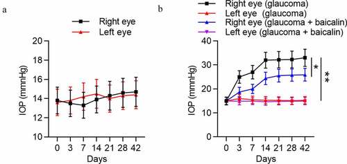
Figure 5. Baicalin increases RGC number and attenuates pathological changes in retinas of glaucoma mice. (a) The structure and morphology of retina of mice in four groups (control, model, Model+NS and Model+BAI) were presented by H&E staining (scale bar = 25 μm). (b) Thickness of GCL and (c) RGCD of mice in four groups (control, model, Model+NS and Model+BAI) were measured. (d) The number of RGCs in retina tissues of four groups (control, model, Model+NS and Model+BAI) was counted. (e) Counting of RGCs using immunoflurescence staining of brain specific homeobox/POU domain protein 3B (Brn3b) in retina tissues of four groups (control, model, Model+NS and Model+BAI). *p < 0.05, ** p < 0.01, *** p < 0.001
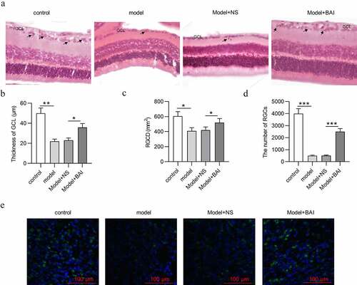
Figure 6. Baicalin inhibits autophagy and activates PI3K/AKT signaling in glaucoma mice. (a) The expression of autophagy-related proteins (LC3-I, LC3-II, Beclin-1 and ATG5) and (b) PI3K/AKT signaling-associated proteins (pAKT, AKT, pPI3K and PI3K) in retinal tissues of four groups (control, model, Model+NS and Model+BAI) was detected by Western blotting. **p < 0.01, *** p < 0.001
Data availability statement
The datasets used during the current study are available from the corresponding author on reasonable request.