Figures & data
Table 1. The sequence of PCR primers used in this study
Table 2. The top 30 significantly downregulated genes from GSE18515 data series using the criteria of P < 0.05 and logFC≤-1.5
Figure 1. The identification of ASF1B as the downstream effector of miR-520d-3p. (a) The protein-protein interaction network of the top 30 significantly downregulated genes of GSE18512 data series. Eight genes were shown in the network. (b) The expression of ASF1B in melanoma (data from GEPIA database). T: tumor; N: normal. (c) The intersection between the target miRNAs of ASF1B predicted by targetscan algorithm and differentially expressed miRNAs (DE-miRNAs) in melanoma from GSE61741 microarray analysis using the criteria of adjusted P < 0.05 and logFC≤-1.5. X
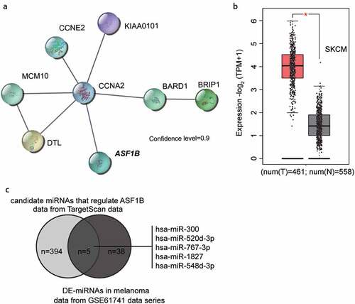
Figure 2. Upregulation of ASF1B in melanoma. (a) RT-qPCR detection of mRNA expression of ASF1B in melanoma tissues (N = 38) and normal tissues (N = 38). (b) RT-qPCR detection of mRNA expression of ASF1B in normal skin cell line (HaCAT) and melanoma cell lines (A375, WM35, A875, and A2058). (c) Measurement of ASF1B protein level in HaCAT, A375, WM35, A875, and A2058 cell lines by western blotting assay. (d) RT-qPCR detection of mRNA expression of ASF1B in A375 and A875 cells transfected with NC, Si-ASF1B and OE-ASF1B. (e) Western blotting detection of protein expression of ASF1B in A375 and A875 cells transfected with NC, Si-ASF1B and OE-ASF1B. *, P < 0.05; **, P < 0.001. CON, blank control; NC, negative control; Si-ASF1B, SiRNA-ASF1B; OE-ASF1B, overexpression-ASF1B
Figure 3. ASF1B facilitated proliferation and adhesion, but repressed cell apoptosis of melanoma cells. (a) Cell viability was detected in A375 and A875 cells transfected with NC, Si-ASF1B and OE-ASF1B by CCK8 assay. (b) Cell proliferation was detected in A375 and A875 cells transfected with NC, Si-ASF1B and OE-ASF1B by BrdU assay. (c) Cell apoptosis was determined in A375 and A875 cells transfected with NC, Si-ASF1B and OE-ASF1B by FITC apoptosis detection kit. (d) Cell apoptosis was determined in A375 and A875 cells transfected with NC, Si-ASF1B and OE-ASF1B by caspase-3 activity assay kit. (e) Cell adhesion was detected in A375 and A875 cells transfected with NC, Si-ASF1B and OE-ASF1B by cell adhesion assay kit. *, P < 0.05; **, P < 0.001. CON, blank control; NC, negative control; Si-ASF1B, SiRNA-ASF1B; OE-ASF1B, overexpression-ASF1B
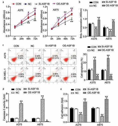
Figure 4. MiR-520d-3p targeting ASF1B inhibited the expression of ASF1B in melanoma. (a) TargetScan analysis showed the predicted binding site of ASF1B 3ʹ-UTR for miR-520d-3p. (b) Dual luciferase assay was performed in pmiRGLO ASF1B-WT and pmiRGLO ASF1B-MUT with treatment of NC or miR-520d-3p mimic. (c) RT-qPCR detection of expression of miR-520d-3p in melanoma tissues and (N = 38) normal tissues (N = 38). (d) Pearson’s correlation analysis of ASF1B mRNA levels and miR-520d-3p in melanoma tissues. (e) RT-qPCR detection of expression of miR-520d-3p in HaCAT, A375 and A875 cells. (f) RT-qPCR detection of expression of miR-520d-3p in A375 and A875 cells transfected with NC, miR-520d-3p inhibitor and miR-520d-3p mimic. (g) Measurement of ASF1B protein level in A375 and A875 cells transfected with NC, miR-520d-3p inhibitor and miR-520d-3p mimic. *, P < 0.05; **, P < 0.001. ASF1B-WT, ASF1B-wild-type; ASF1B-MUT, ASF1B-mutunt; CON, blank control; NC, negative control; inhibitor, miR-520d-3p inhibitor; mimic, miR-520d-3p mimic
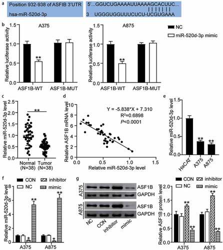
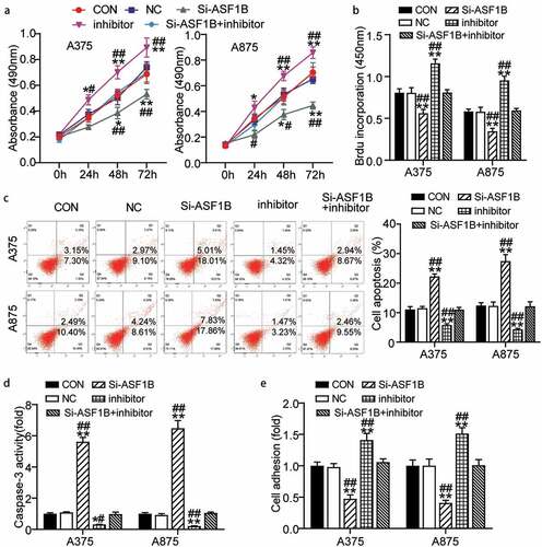
Figure 5. MiR-520d-3p hampered melanoma cell progression by inhibiting ASF1B. (a) Cell viability was detected in A375 and A875 cells transfected with NC, Si-ASF1B, miR-520d-3p inhibitor and Si-ASF1B+miR-520d-3p inhibitor by CCK8 assay. (b) Cell proliferation was detected in A375 and A875 cells transfected with NC, Si-ASF1B, miR-520d-3p inhibitor and Si-ASF1B+miR-520d-3p inhibitor by BrdU assay. (c) Cell apoptosis was determined in A375 and A875 cells transfected with NC, Si-ASF1B, miR-520d-3p inhibitor and Si-ASF1B+miR-520d-3p inhibitor by FITC apoptosis detection kit. (d) Cell apoptosis was determined in A375 and A875 cells transfected with NC, Si-ASF1B, miR-520d-3p inhibitor and Si-ASF1B+miR-520d-3p inhibitor by caspase-3 activity assay kit. (e) Cell adhesion was detected in A375 and A875 cells transfected with NC, Si-ASF1B, miR-520d-3p inhibitor and Si-ASF1B+miR-520d-3p inhibitor by cell adhesion assay kit. *, P < 0.05; **, P < 0.001. CON, blank control; NC, negative control; Si-ASF1B, SiRNA-ASF1B; inhibitor, miR-520d-3p inhibitor