Figures & data
Figure 1. Expression of circANKS1B and TGF-β1 in OSCC tissues and cells. (a) The genomic loci of circANKS1B (has_ circ_0007294). (b, c) qRT-PCR assay was performed to detect the expression of circANKS1B (b) and TGF-β1 (c) in OSCC tissues. (d) Pearson’s correlation analysis was utilized to elucidate the association between circANKS1B and TGF-β1. (e) Expression of circANKS1B in OSCC cells and normal oral keratinocyte HOK cells. *P < 0.05
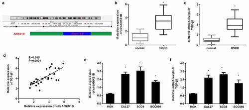
Figure 2. circANKS1B positively regulates TGF-β1 expression in OSCC cell lines. (a) OSCC cells (CAL27 and SCC9 cells) were transfected with the recombinant circANKS1B plasmids. The expression of circANKS1B was then determined. (b) Effects of circANKS1B overexpression on the mRNA levels of TGF-β1. (c) The expression of circANKS1B was detected in cells transfected with si-circANKS1B. (d) Expression of TGF-β1 after si-circANKS1B transfection. (e-h) The expression of TGF-β1 was analyzed by western blotting (e and g) and ELISA (f, h) in cells transfected with circANKS1B vectors or si-circANKS1B. *P < 0.05
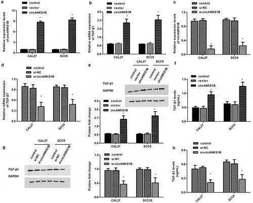
Figure 3. Effects of circANKS1B knockdown on OSCC cell metastatic potential. (a, b) After transfection with si-circANKS1B, cell invasion was evaluated by MTT assay in CAL27 (a) and SCC9 (b) cells. (c, d) Effects of circANKS1B inhibition on cell migration. (e, f) Protein expression of EMT marker E-cadherin and N-cadherin in CAL27 and SCC9 cells. The corresponding bands were quantified by ImageJ software. *P < 0.05
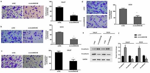
Figure 4. Silencing cicrANKS1B sensitizes OSCC cells to cisplatin. (a, b) After transfection with si-circANKS1B in cells under cisplatin exposure, cell viability was then assessed. (c) The subsequent effects on cell apoptosis were analyzed by Annexin V-FITC/PI staining. (d, e) The activity of caspase-3 was detected. *P < 0.05
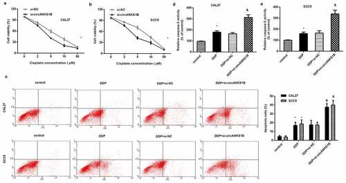
Figure 5. circANKS1B acts as a sponge for miR-515-5p to regulate TGF-β1expression. (a) The potential binding site between circANKS1B (circ_0007294) and miR-515-5p. (b) The expression of miR-515-5p in normal and OSCC tissues. (c) The expression of miR-515-5p in OSCC cells. (d) After co-transfection with circANKS1B-wt/mut luciferase reporter plasmids and miR-515-5p mimics, the luciferase activity was determined. (e) The relationship among circANKS1B and miR-515-5p was determined by RIP assay. (f) Starbase 3.0 was applied to predict the binding site between miR-515-5p and TGF-β1. (g-i) The relationship among miR-515-5p and TGF-β1 was analyzed by qRT-PCR and luciferase activity assay
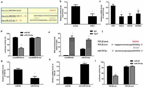
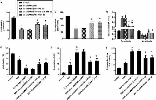
Figure 6. circANKS1B regulates the metastatic potential and cisplatin resistance by sponging miR-515-5p/ TGF-β1. (a-c) CAL27 cells were treated with si-circANKS1B, anti-NC, anti-miR-515-5p or TGF-β1. Then, cell invasion (a), migration (b), and EMT marker expression (E-cadherin and N-cadherin) (c) were analyzed. *P < 0.05 vs. control group, &P < 0.05 vs. si-circANKS1B group. (d-f) Cells under si-circANKS1B, anti-NC, anti-miR-515-5p or TGF-β1 were exposed to cisplatin. Then, cell viability (d), apoptosis (e) and caspase-3 activity (f) were determined. *P < 0.05 vs. control group, &P < 0.05 vs. DDP-treated group, $P < 0.05 vs. DDP and si-circANKS1B-treated group