Figures & data
Figure 1. DHI improves DN.
(a) Blood glucose test every 4 weeks; (b) weight check every 4 weeks; (c) creatinine measurement at 24 weeks of age; (d) urea nitrogen detected at 24 weeks of age; (e-g) inflammatory markers IL-6, IFN-γ and TNF-α contents in serum of db/db mice; (h) the pathological condition of renal tissues detected via HE staining; (i) the renal tissue apoptosis detected via TUNEL staining. The data were expressed as mean ± SEM, (a&b) n = 10; C-I, n = 5; * P < 0.05, versus db/m; # P < 0.05, versus db/db control.
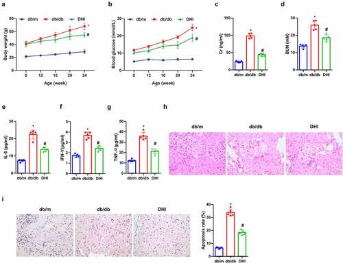
Figure 2. Overexpression of miR-30d-5p alleviates renal dysfunction in db/db mice.
(a) The relative expression of miR-30d-5p in renal tissues detected via Real-time PCR; (b) blood glucose test every 4 weeks; (c) weight check every 4 weeks; (d) creatinine measurement at 24 weeks of age; (e) urea nitrogen detected at 24 weeks of age; (f-h) inflammatory markers IL-6, IFN-γ and TNF-α contents in serum of db/db mice; (i) the pathological condition of renal tissues detected via HE staining; (j) the renal tissue apoptosis detected via TUNEL staining. The data were expressed as mean ± SEM, (b&c) n = 10; (a&d–j), n = 5; + P < 0.05, versus DHI + NC.
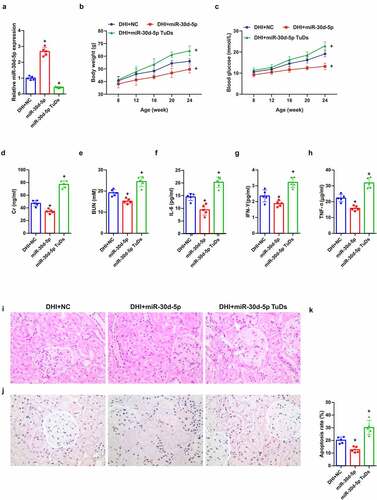
Figure 3. DHI regulates the miR-30d-5p-JAK1 axis in vivo.
(a/b) The expression of miR-30d-5p and JAK1 in renal tissues detected by qPCR; (c/d) the protein expression of JAK1 in renal tissues detected via Western Blot. The data were expressed as mean ± SEM, n = 5; * P < 0.05, versus db/m; # P < 0.05, versus db/db control.
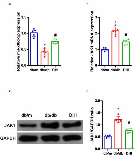
Figure 4. DHI treatment mitigates in vitro cell damage in DR.
(a/b) Cell viability detected by MTT assay; (c) cell proliferation detected by plate cloning; (d) apoptosis detected via Flow cytometry; (e) cell migration detected via Transwell assay; (f) the ability of cytological tubule formation tested via Tubule formation assay. The data were expressed as mean ± SEM, N = 3; * P < 0.05, versus Blank; # P < 0.05, versus HG.
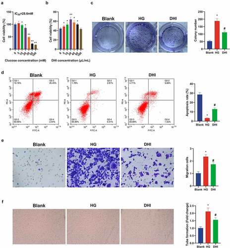
Figure 5. DHI upregulated miR-30d-5p and targeting JAK1 in vitro.
(a/b) The expression of miR-30d-5p and JAK1 in ARPE-19 cells detected by qPCR; (c) the protein expression of JAK1 in ARPE-19 cells detected via Western Blot; (d) the binding sites of miR-30d-5p and JAK1 predicted via Bioinformatics website (TargetScan); (e/f) the expression of miR-30d-5p and JAK1 after upregulation of miR-30d-5p detected by qPCR. (c) The protein expression of JAK1 after up-regulation of miR-30d-5p detected via Western Blot. The data were expressed as mean ± SEM, N = 3; * P < 0.05, versus Blank; # P < 0.05, versus HG; + P < 0.05, versus miR-NC.
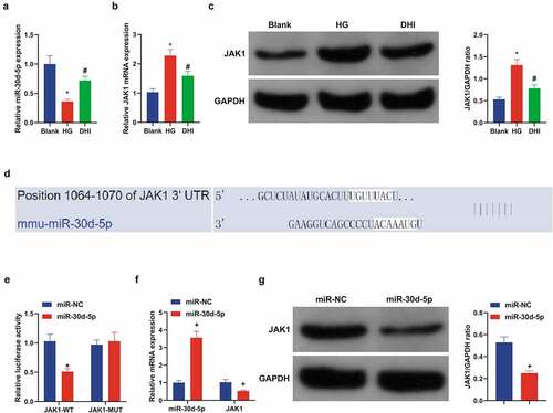
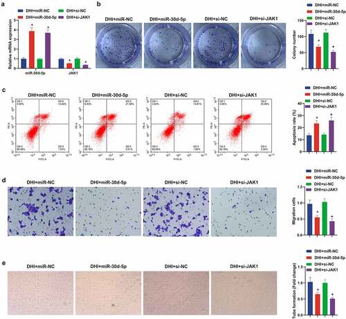
Figure 6. Down-regulating miR-30d-5p or up-regulating JAK1 can facilitate the development of DR model in vitro and remove the protective effect of DHI in vitro.
(a) The transfection efficiency verified via qPCR; (b) cell proliferation detected by plate cloning; (c) apoptosis detected via Flow cytometry; (d) cell migration detected via Transwell assay; (e) the ability of cytological tubule formation tested via Tubule formation assay. The data were expressed as mean ± SEM, N = 3; ^ P < 0.05, versus in-NC; + P < 0.05, versus oe-NC.