Figures & data
Table 1. Sequences of primers used for reverse transcription-quantitative PCR
Figure 1. MiR-191-5p is downregulated in the hippocampal tissues of APP/PS1 mice. (a-b) Pathological changes of the hippocampus in C57BL/6 J mice and APP/PS1 mice were assessed by H&E staining and Nissl staining. (c) Aβ-positive granules in the hippocampal tissues of C57BL/6 J mice and APP/PS1 mice were determined by immunohistochemical staining. (b) MiR-191-5p expression in the hippocampal tissues of C57BL/6 J mice (n = 8) and APP/PS1 mice (n = 24) was detected by RT-qPCR. ***p < 0.001
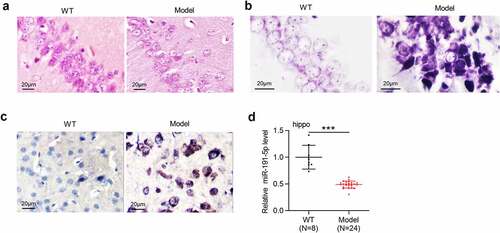
Figure 2. MiR-191-5p overexpression alleviates Aβ1-42-induced microglial cell injury. (a) MiR-191-5p expression in microglia treated with or without Aβ1-42, treated with Aβ1-42 and transfected with NC mimics, treated with Aβ1-42 and transfected with miR-191-5p mimics was measured by RT-qPCR. (b) Viability of microglial cells in the above four groups (Con, Aβ1-42, Aβ1-42+ NC mimics, Aβ1-42+ miR-191-5p) was assessed by CCK-8 assays. (c-d) Apoptosis of microglial cells in the above four groups was detected by flow cytometry analyses. (e-f) Western blotting was performed to assess protein levels of BACE1 and Tau-5 in microglial cells in the above four groups. *p < 0.05, **p < 0.01, ***p < 0.001
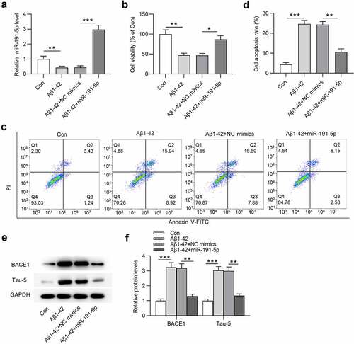
Figure 3. MiR-191-5p targets Map3k12. (a) Potential target genes (Taf5, Neurl4, Tmod2, Chmp5, Sall1, Tjp1, Map3k12 and Wiz) of miR-191-5p were predicted by miRDB with the screening condition of target score > 85. (b) The mRNA levels of potential target genes (Taf5, Neurl4, Tmod2, Chmp5, Sall1, Tjp1, Map3k12 and Wiz) of miR-191-5p in microglial cells after transfection of NC mimics or miR-191-5p mimics were examined by RT-qPCR. (c) Map3k12 protein level in microglial cells after transfection of NC mimics or miR-191-5p mimics was detected by Western blotting. (d) A binding site between miR-191-5p and Map3k12 3ʹ-UTR was predicted Targetscan. (e) Luciferase reporter assay was performed to confirm the binding relationship between miR-191-5p and Map3k12. **p < 0.01
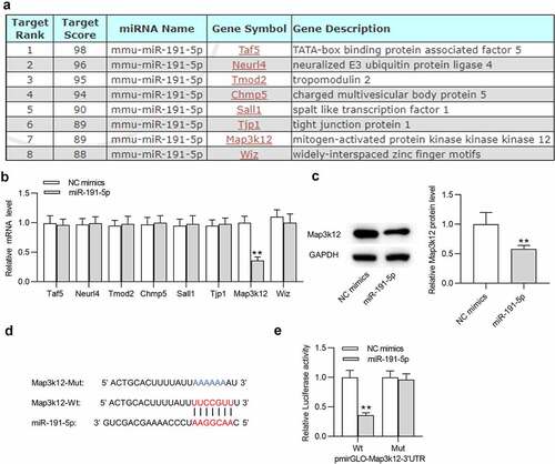
Figure 4. MiR-191-5p alleviates Aβ1-42-induced microglial cell injury by downregulating Map3k12. (a-b) Map3k12 protein level in Aβ1-42-treated microglia transfected with NC mimics, miR-191-5p mimics, or cotransfected with miR-191-5p mimics and pcDNA3.1/Map3k12 was assessed by Western blotting. (c) Viability of Aβ1-42-stimulated microglia in above three groups (NC mimics, miR-191-5p, miR-191-5p+Map3k12) was measured by CCK-8 assays. (d-e) Apoptosis of Aβ1-42-treated microglial cells in above three groups was detected by flow cytometry analyses. (f-g) The protein levels of BACE1 and Tau-5 in Aβ1-42-treated microglia from the three groups were evaluated by Western blotting. *p < 0.05, **p < 0.01, ***p < 0.001
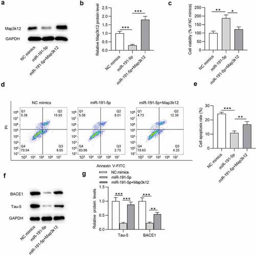
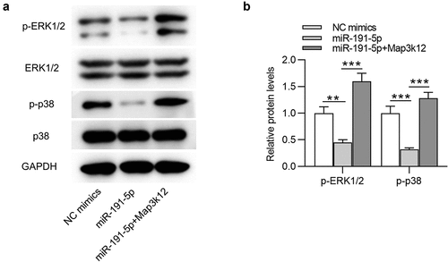
Figure 5. MiR-191-5p inactivates the MAPK signaling by targeting Map3k12. (a-b) Western blotting was performed to quantify protein levels of MAPK signaling-associated factors (ERK1/2, p-ERK1/2, p38, p-p38) in Aβ1-42-treated microglia transfected with NC mimics, miR-191-5p mimics or miR-191-5p mimics + pcDNA3.1/Map3k12. **p < 0.01, ***p < 0.001