Figures & data
Table 1. Clinical characteristics of AS patients
Figure 1. TMAO promotes the development of AS in vivo.
A: Oil red O assay showed that TMAO increased atherosclerotic plaques and lipid contents. B: TMAO facilitated the increase in lesion area induced by HF. C: The levels of TMAO in vivo was determined using ELISA. **P < 0.01.
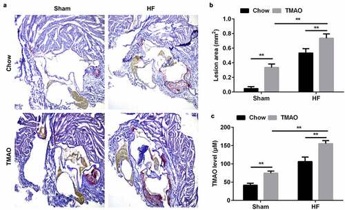
Figure 2. TMAO was increased in AS patients.
A: The plasma concentration of TMAO in AS patients determined using ELISA. B: Cell viability was determined using CCK-8 assay. *P < 0.05, **P < 0.01.
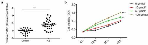
Figure 3. TMAO increases the level of NEAT1 in AS.
A: The expression of NEAT1 in AS patients determined using qRT-PCR. B: The expression of NEAT1 analyzed using AUC curve. C: NEAT1 was overexpressed in cells treated with TMAO. **P < 0.01.
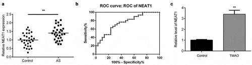
Figure 4. Knockdown of NEAT1 modulates the cell proliferation and apoptosis of HUVECs.
A: The expression of NEAT1 determined using qRT-PCR. B: The cell viability of HUVECs detected using CCK-8. C: The colony numbers determined using colony formation assay. D: The proliferation of HUVECs determined by EdU assay. E: The apopotsis of HUVECs detected using flow cytometry assay. **P < 0.01.
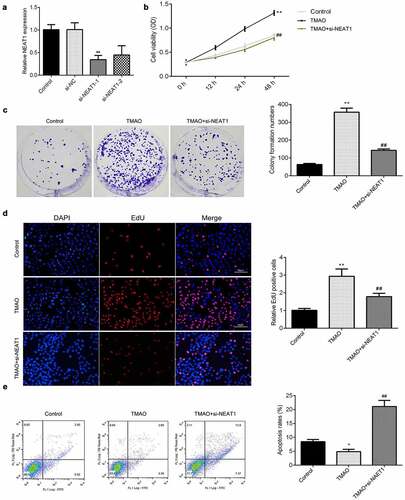
Figure 5. STAT3 modulates the progression of AS.
A: The mRNA level of STAT3 in AS patients determined using qRT-PCR. B: The protein level of STAT3 in AS patients determined using Western blot. **P < 0.01.
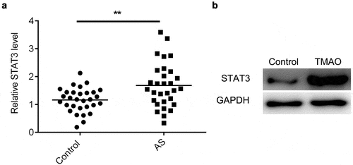
Figure 6. NEAT1 regulates the expression of STAT3 via sponging miR-370-3p.
A: The binding sites between miR-370-3p and NEAT1 verified using luciferase activity assay. B: The binding sites between miR-370-3p and STAT3 verified using luciferase activity assay. C: The protein level of STAT3 detected using Western blot. D: Quantification of C. E: The protein level of STAT3 detected using Western blot. F: Quantification of E. G: The expression of miR-370-3p in AS patients determined using qRT-PCR. H: The correlation analysis performed using Pearson method. I: The correlation analysis performed using Pearson method. **P < 0.01.
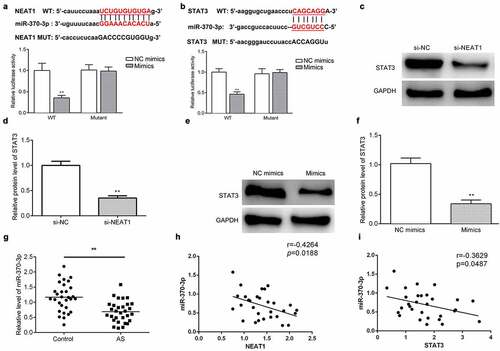
Figure 7. NEAT1 regulates the proliferation and apoptosis of HUVECs via sponging miR-370-3p.
A: The expression of miR-370-3p determined using qRT-PCR. B: The cell viability of HUVECs detected using CCK-8. C: The colony numbers determined using colony formation assay. D: The proliferation of HUVECs determined by EdU assay. E: The apopotsis of HUVECs detected using flow cytometry assay. **P < 0.01, ##P < 0.01.
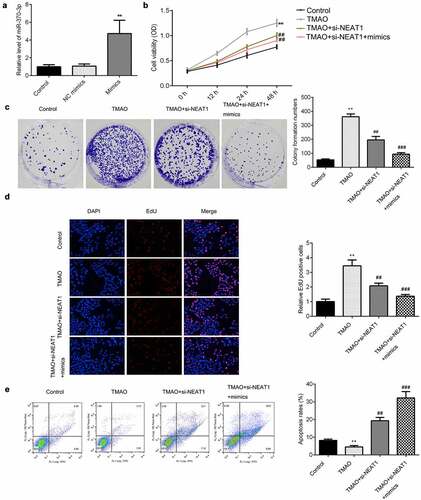
Figure 8. NEAT1 regulates the progression of AS via regulating STAT3.
A: The expression of STAT3 determined using qRT-PCR. B: The cell viability of HUVECs detected using CCK-8. C: The colony numbers determined using colony formation assay. D: The proliferation of HUVECs determined by EdU assay. E: The apopotsis of HUVECs detected using flow cytometry assay. **P < 0.01, ##P < 0.01.

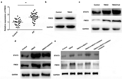
Figure 9. FMO3 is a potential target of STAT3 pathways.
A: The mRNA level of FMO3 detected using qRT-PCR. B: The protein level of FMO3 determined using Western blot. C: The protein level of FMO3 determined using Western blot. D: The protein expression of STAT3 and FMO3 detected using Western blot. E: The protein expression of STAT3 and FMO3 detected using Western blot. **P < 0.01.