Figures & data
Table 1. Primer sequence
Figure 1. Elevation of circ-PSEN1 by HG treatment in ARPE19 cells. (a) RT-qPCR determined expression levels of circ-PSEN1 in the ARPE19 cells treated with normal level glucose and HG for 48 hours. (b) The RNA levels of circ-PSEN1 and linear PSEN1 were measured by RT-qPCR after RNase R was incubated with the total cellular RNA. Mock treated cells were used as the negative control. (c) Relative RNA levels of circ-PSEN1 and PSEN1 were recorded 0, 4, 8, 12, 24 h after the cells were treated by actinomycin D. Data are representative of three experiments. **P < 0.01; ***P < 0.001. HG, high glucose; ARPE19, adult retinal pigment epithelial cell line-19; Act D, actinomycin D
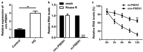
Figure 2. Si-circ-PSEN1 regulates contents of GSH, Fe2+, and MDA in the HG-treated ARPE19 cells. (a) The expression levels of circ-PSEN1 when the ARPE19 cells were transfected with si-circ-PSEN1 1# and si-circ-PSEN1 2#. (b) GSH, (c) MDA, and (d) Fe2+ contents were examined in the HG-treated cells 48 h after transfection with si-circ-PSEN1 using the corresponding kits. Data are representative of three experiments; *P < 0.05; **P < 0.01. HG, high glucose; ARPE19, adult retinal pigment epithelial cell line-19; GSH, glutathione; MDA, malondialdehyde
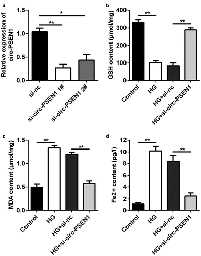
Figure 3. Downregulation of circ-PSEN1 alleviates ferroptosis of HG-induced ARPE19 cells. (a) The MTT assay was used to assess the cell survival rate of the cells treated with HG and si-circ-PSEN1 for 24 h. (b) PI positive cells were photographed and counted by fluorescence microscopy. DAPI was used for counter staining. The ARPE19 cells were treated with HG and si-circ-PSEN1 for 24 h. (c) TUNEL positive cells were captured and counted by fluorescence microscopy. The ARPE19 cells were treated with HG and si-circ-PSEN1 for 24 h. (d) The protein expressions of GPX4, SLC7A11, and TFR1 were detected by Western blotting 24 h after the indicated treatments. Data are representative of three experiments; **P < 0.01. HG, high glucose; ARPE19, adult retinal pigment epithelial cell line-19; PI, propidium iodide; DAPI, 4′,6-diamidino-2-phenylindole; TUNEL, TdT mediated dUTP Nick End Labeling
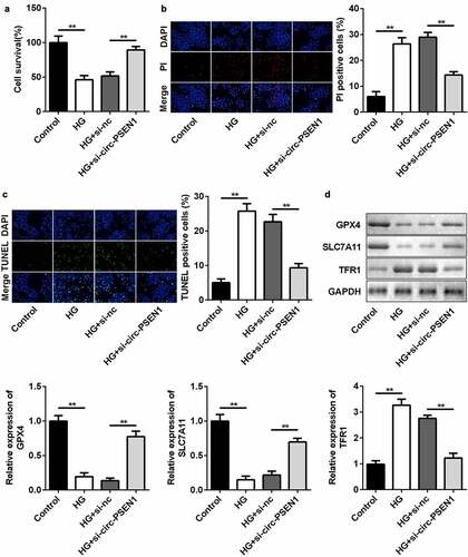
Figure 4. miR-200b-3p is the target of circ-PSEN1. (a) The sequences of circ-PSEN1 WT, circ-PSEN1 MUT, and miR-200b-3p. (b) Luciferase activities of the ARPE19 cells were analyzed 24 h after the indicated treatments. (c) The levels of circ-PSEN1 enriched by negative control and biotin-labeled miR-200b-3p. (d) The expression levels of miR-200b-3p of the cells transfected with circ-PSEN1, si-circ-PSEN1, or their control. (e) The expression level of miR-200b-3p in the HG-treated ARPE19 cells. Data are representative of three experiments; **P < 0.01. HG, high glucose; ARPE19, adult retinal pigment epithelial cell line-19; WT, wild type; MUT, mutant type
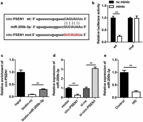
Figure 5. Inhibition of miR-200b-3p abrogates the effects of si-circ-PSEN1 on ferroptosis. (a) The expression level of miR-200b-3p in ARPE19 cells transfected with miR-200b-3p inhibitor. (b) GSH contents, (c) MDA contents, and (d) Fe2+ in HG-treated cells 48 h after transfection. (e) The MTT assay was used to assess the cell survival rate of the cells treated with HG and the indicated plasmids after 24 h. (f) PI positive cells and (g) TUNEL positive cells were photographed and counted by fluorescence microscopy. (h) Protein expressions of GPX4, SLC7A11, and TFR1 detected by Western blotting assay 24 h after the indicated treatments. Data are representative of three experiments; *P < 0.05; **P < 0.01. HG, high glucose; ARPE19, adult retinal pigment epithelial cell line-19; GSH, glutathione; MDA, malondialdehyde; PI, propidium iodide; DAPI, 4ʹ,6-diamidino-2-phenylindole; TUNEL, TdT mediated dUTP Nick End Labeling
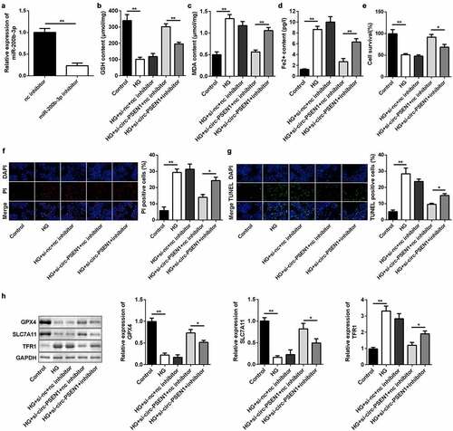
Figure 6. CFL2 is the target gene of miR-200b-3p. (a) The sequences of CFL2 WT, CFL2 MUT and miR-200b-3p. (b) Luciferase activities of the ARPE19 cells were analyzed 24 h after the indicated treatments. (c) The levels of CFL2 enriched by negative control and biotin-labeled miR-200b-3p. (d) The expression levels of CFL2 of the cells transfected with miR-200b-3p mimic, miR-200b-3p inhibitor, or their control. (e) The expression level of CFL2 in the HG-treated ARPE19 cells. Data are representative of three experiments; **P < 0.01. CFL2, cofilin-2; HG, high glucose; ARPE19, adult retinal pigment epithelial cell line-19; WT, wild type; MUT, mutant type
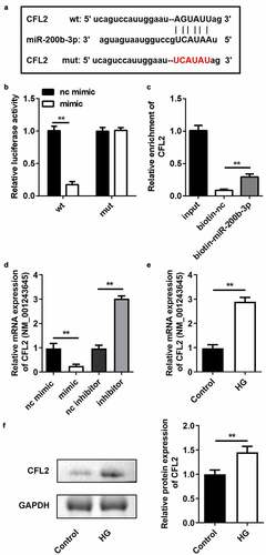
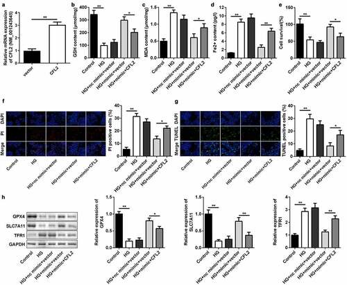
Figure 7. CFL2 reverses the effects of miR-200b-3p on cell ferroptosis. (a) The expression level of CFL2 in the ARPE19 cells transfected with CFL2/pcDNA3.1. (b) GSH, (c) MDA, and (d) Fe2+ contents were examined in HG-treated cells 48 h after transfection. (e) The MTT assay was used to assess the cell survival rate of the cells treated with HG and the indicated vectors for 24 h. (f) PI positive cells and (g) TUNEL positive cells were photographed and counted by fluorescence microscopy. (h) Protein expressions of GPX4, SLC7A11, and TFR1 were detected by Western blotting 24 h after the cells underwent the indicated treatments. The mRNA expression levels of their genes were detected by RT-qPCR. Data are representative of three experiments; *P < 0.05; **P < 0.01. CFL2, cofilin-2; HG, high glucose; ARPE19, adult retinal pigment epithelial cell line-19; GSH, glutathione; MDA, malondialdehyde; PI, propidium iodide; DAPI, 4′,6-diamidino-2-phenylindole; TUNEL, TdT mediated dUTP Nick End Labeling