
Figures & data
Table 1. Correlationship between COAD patient characteristics and KRT 17 level (*P < 0.05). – – TCGA
Figure 1. KRT17 upregulation in COAD. (a, b) Data from Oncomine and TCGA databases shows that KRT17 is upregulated in COAD as compared to the normal control tissues. (c) KRT17 expression in 20 paired COAD and matched adjacent normal tissues from TCGA database; P-values are shown (n = 20). (d) RT-PCR analysis of KRT17 expression in 25 COAD tissues; ΔCT value is inversely proportional to relative expression. (e) KRT17 expression is related to the N stage (*P < 0.05 **P < 0.01 ***P < 0.001). (f) Immunohistochemical staining for KRT17 in COAD and normal tissues
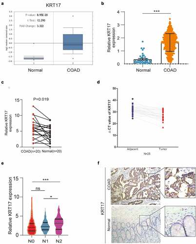
Figure 2. KRT17 promotes the migration and invasion of colon cancer cells. (a, b) Western blotting and RT-PCR analyses of KRT17 expression in seven colon cancer cell lines; mean ± SEM are displayed (n = 3). (c) WB analysis for the efficiency of si-KRT17 and si-Control transfection in SW116 cells; overexpression plasmid and empty control vector transfection in LOVO cells. (d) Wound healing assay for cell migration in SW116 and LOVO cells; the images of wound closure are shown for the indicated number of hours after scratching (0, 24 h). (e) The quantification for wound healing assay. (f) Transwell assays to examine the potential migration and invasion of transfected cells. (g) The quantification of (F). (h) intravital imaging assay for cell metastasis in SW116 KRT17 Con and SW116 KRT17 si cells
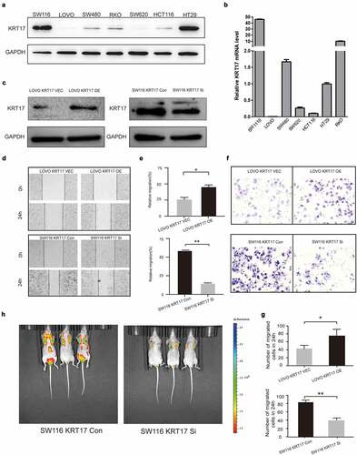
Figure 3. Overexpression of KRT17 induces angiogenesis in vivo. (a) Immunohistochemical staining for CD31 in COAD and normal tissues. (b) The effect of KRT17 expression on the tube formation ability of HUVEC by tube formation assay; mean ± SEM are displayed (n = 3). (c) Matrigel angiogenesis assay reflects the effect of KRT17 expression on neovascularization in comparison with the control group. (d) H&E staining of Matrigel sections and the number of MVDs; mean ± SEM are displayed (n = 3). (e) Images of the blood vessels of chick embryos; bar chart demonstrates the relative number of blood vessels; data are presented for at least three independent experiments (*P < 0.05 **P < 0.01 according to the two-tailed Student’s t-test)
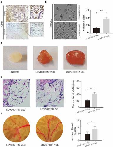
Figure 4. The relationship between KRT17 and WNT signaling pathway. (a) The result above meets the criteria of FDR<0.25, P-value < 0.05; high expression of KRT17 is significantly related to the WNT signaling pathway. (b, c) Western blotting and RT-PCR analyses of KRT17 expression at different concentrations of Wnt3a (0, 2, and 10 μM) treatment for 24 hours. (d) Protein level expression of Wnt/β-catenin pathway members, β-catenin and KRT17, in SW116 KRT17 Con cells and SW116 KRT17 si cells with or without Wnt3a (2 μM) pretreatment
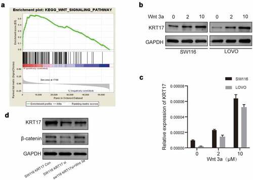
Figure 5. Correlation between KRT17 and APC genes. (a) Using the STRING online database PPI network of the KRT17 was constructed. (b) Co-immunoprecipitation shows that KRT17 interacts with APC in human COAD cell lines. Total cell lysate from SW116 cells was immunoprecipitated with anti-APC and anti-KRT17 antibodies. (c) Protein level expression of KRT17, β-catenin, and APC in SW116 cells. SW116 APC OE cells with or without Wnt3a (2 μM) pretreatment. (d, e) Wound healing and Transwell assays show that APC overexpression inhibits the migration and invasion of SW116 cells; mean ± SEM are displayed (n = 3) (*P < 0.05 **P < 0.01 according to the two-tailed Student’s t-test)
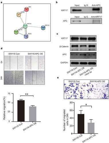
Table 2. Correlationship between the clinicopathological features of COAD patients and the level of KRT17 (*P < 0.05). – – Affiliated Hospital of Nantong University
Supplemental Material
Download Zip (25.6 MB)Data availability statement
The raw data supporting the conclusions of this manuscript will be made available by the authors, without undue reservation, upon request to any qualified researcher. The data sources in the manuscript are as follows:TCGA:https://portal.gdc.cancer.gov/;string:https://cn.string-db.org/;GSEA:http://www.gsea-msigdb.org/gsea/index.js