Figures & data
Figure 1. The expression level of circRbms1 in the I/R mice and H2O2 treated H9c2 cells. (a) The level of circRbms1 in the I/R mice was detectedd by RT-PCR. (b) The level of circRbms1 H2O2 treated H9c2 cells was detectedd by RT-PCR. Each experiment was repeated three times. **P < 0.01.
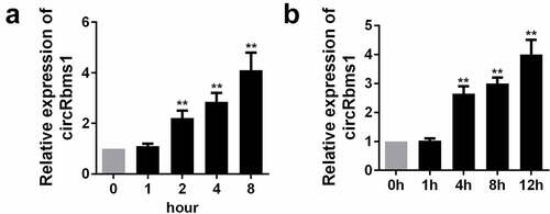
Figure 2. Silencing circRbms1 can attenuate apoptosis and oxidative stress injury of H9c2 cells induced by H2O2. (a) circRbms1 expression was determined by RT-PCR. (b, c) Apoptosis of H9c2 cells was detected using flow cytometry. (d) The apoptosis was detected by TUNEL assays. (e) Reactive oxygen species was assessed. Each experiment was repeated three times. **P < 0.01.
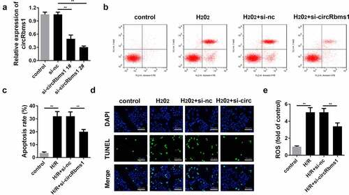
Figure 3. Silencing circRbms1 inhibits I/R injury in mice. (a) circRbms1 and miR-92 expression was determined by RT-PCR. (b) The AMI was analyzed using TTC staining. (c) The histopathology examination of myocardial tissues was performed with HE staining. (d) Primary cardiomyocyte apoptosis was evaluated by TUNEL assay. Each experiment was repeated three times. **P < 0.01.
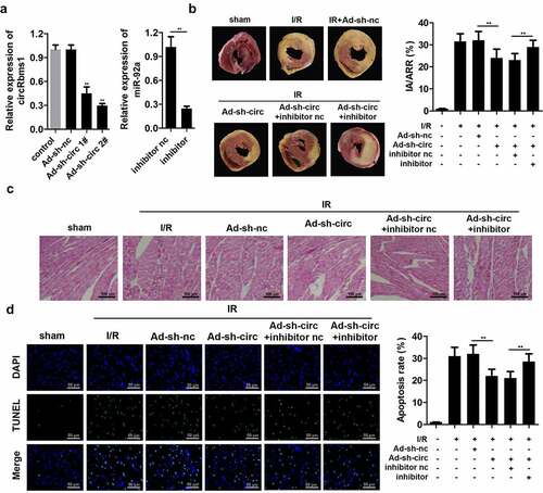
Figure 4. circRbms1 sponges miR-92a. (a) The binding sites of circRbms1 and miR-92a. (b) circRbms1 negatively mediates miR-92a expression. The binding sites were confirmed by luciferase report analysis (c) and RNA pull-down assay (d). Each experiment was repeated three times. **P < 0.01.
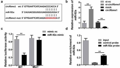
Figure 5. MiR-92a inhibition reverses the effect of circRbms1 knockdown. (a) miR92a expression was determined by RT-PCR. (b-d) Apoptosis rates of H9c2 cells were evaluated by flow cytometry and TUNEL staining. (e) The reactive oxygen species in H9c2 cells was detected. Each experiment was repeated three times. **P < 0.01.
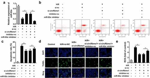
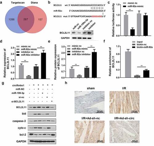
Figure 6. MiR-92a inhibits cardiomyocyte apoptosis by regulating BCL2L11. (a) The TargetScan database and Diana database predicted target genes of miR-92a. (b) The binding sites between miR-92a and BCL2L11. (c) The binding sites were confirmed by luciferase report analysis. (d) BCL2L11 expression was determined by RT-PCR. (e) BCL2L11 protein expression was evaluated by Western blot. (f) The binding sites were confirmed by RNA pull-down assay. (g) Protein expression was detected using Western blot. (h) BCL2L11 protein expression was evaluated by IHC staining. Each experiment was repeated three times. **p < 0.01.