
Figures & data
Figure 1. GJ relieves I/R rat neuronal injury. (a) GJ chemical structure. (b) Experimental protocol schematic diagram. (c-f) MCAO/R rats were treated with GJ at indicated doses (n = 6). (c) Rat neurological deficit scores were measured after 24 h of reperfusion. (d) TC staining of cerebral infarction in rats. (e) Rat brain water content was calculated after 24 h of reperfusion. (f) TC staining of cerebral infarction after reperfusion in the rats. ** P < 0.01, # P < 0.05, ## P < 0.01. Data are presented as mean ± SD.
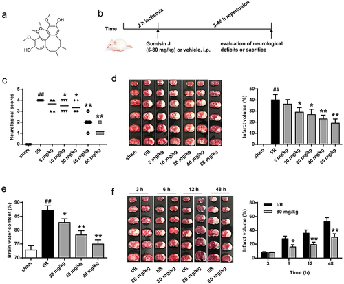
Figure 2. GJ attenuates the neurological loss of hippocampus in I/R rats. MCAO/R rat model was constructed and treated with GJ at indicated doses (n = 6). Hematoxylin & Eosin staining of hippocampus neurons in the rats (200×). * P < 0.05, ** P < 0.01, ## P < 0.01. Data are presented as mean ± SD.
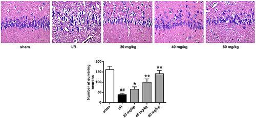
Figure 3. GJ represses apoptosis of ischemic tissues in I/R rats. MCAO/R rats were constructed and treated with GJ at indicated doses (n = 6). The apoptosis of cerebral cortex tissues was analyzed by TUNEL staining. * P < 0.05, ** P < 0.01, ## P < 0.01. Data are presented as mean ± SD.
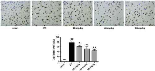
Figure 4. GJ regulates apoptotic proteins in I/R rat ischemic tissues. (a-d) MCAO/R rats were constructed and treated with GJ at indicated doses (n = 6). β-actin, cleaved caspase-3, Bax, and Bcl-XL levels was analyzed by Western blot. * P < 0.05, ** P < 0.01, ## P < 0.01. Data are presented as mean ± SD.
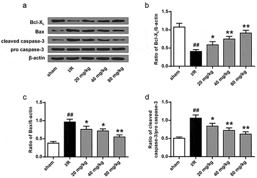
Figure 5. GJ inhibits inflammation in I/R rats. (a-d) MCAO/R rats were constructed and treated with GJ at indicated doses (n = 6). (a-c) NF-κB (p-p65), COX-2, and β-actin expression was measured. (d) NO levels were analyzed in rats. * P < 0.05, ** P < 0.01, ## P < 0.01. Data are presented as mean ± SD.
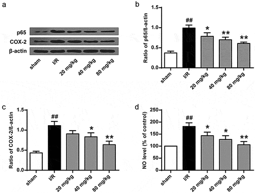
Figure 6. GJ inhibits oxidative stress in I/R rats. (a-f) MCAO/R rats were constructed and treated with GJ at indicated doses (n = 6). (a-c) Nucleus Nrf2, Lamin B1, HO-1, and β-actin expression was measured by Western blot. (d-f) SOD and GSH-Px activities and GSH content were analyzed. * P < 0.05, ** P < 0.01, ## P < 0.01. Data are presented as mean ± SD.
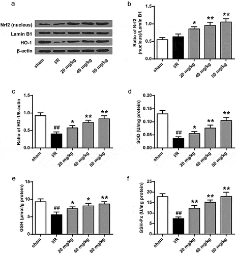
Figure 7. GJ decreases lipid peroxidation in I/R rats. MCAO/R rats were constructed and treated with GJ at indicated doses (n = 6). MDA levels were analyzed in rats. * P < 0.05, ** P < 0.01, ## P < 0.01. Data are presented as mean ± SD.
