
Figures & data
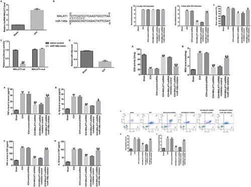
Figure 1. Long noncoding RNA MALAT1 was upregulated in intracerebral hemorrhage rats targeted miR-146a and miR-146a was downregulated. (a)The expression levels were detected using quantitative real-time PCR (qRT-PCR). (b) Binding sites between MALAT1 and miR-146a. (c) Dual-luciferase analysis proved that the miR-146a mimic repressed the luciferase activity of WT‑MALAT1, while no obvious effects on MUT‑MALAT1 activity. (d) The qRT-PCR assay was used to check miR-146a levels in intracerebral hemorrhage rats. Results are displayed as mean ± standard deviation, **p < 0.01, vs. sham; ##p < 0.01 vs. mimic control.
Figure 2. Inhibition of long noncoding RNA MALAT1 reduced neurological damage and brain edema in intracerebral hemorrhage rats. After 1 h of intracerebral hemorrhage (ICH) induction, control-shRNA, lncRNA MALAT1-shRNA, MALAT1-shRNA+inhibitor control, or MALAT1-shRNA+miR-146a inhibitor were transfected into ICH rats. (a) MALAT1 levels in different groups. (b) Expression of miR-146a in different groups. Neurobehavioral scores were assessed using the mNSS test in various groups after ICH for 1 h (c) and 3 d (d). (e) Brain water content was investigated using the wet/dry method in all groups. Data are displayed as means ± standard deviation, **p < 0.01 vs. sham; #, ##p < 0.05, 0.001 vs. ICH+control-shRNA; &, &&p < 0.05, 0.01 vs. ICH+MALAT1-shRNA+inhibitor control.
Figure 3. Downregulation of long noncoding RNA MALAT1 inhibited the pro‑inflammatory cytokine levels in intracerebral hemorrhage rats. ELISA was employed to determine the TNF-α and IL-1β levels in the serum and cerebrospinal fluid in rats of sham, ICH, ICH+control-shRNA, ICH+MALAT1-shRNA, ICH+MALAT1-shRNA+inhibitor control, and ICH+MALAT1-shRNA+miR-146a inhibitor groups. Data are expressed as the mean ± SD. **p < 0.01 vs. Sham; ##p < 0.001 vs. ICH+control-shRNA; &, &&p < 0.05, 0.01 vs. ICH+MALAT1-shRNA+inhibitor control.
Figure 4. Downregulation of long noncoding RNA MALAT1 suppressed oxidative stress in intracerebral hemorrhage rats.
Figure 5. Inhibition of long noncoding RNA MALAT1 inhibited neuronal apoptosis in intracerebral hemorrhage rats.
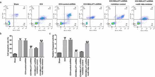
Figure 6. Downregulation of long noncoding RNA MALAT1 suppressed the NF‑κB pathway in intracerebral hemorrhage rats.
Data Availability Statement
The datasets used and/or analyzed during the current study are available from the corresponding author upon reasonable request.