Figures & data
Table 1. Primers and sequences used in the present research
Figure 1. Knockdown of LINC00665 suppressed cell proliferation in breast cancer cells.
(A) The expression of LINC00665 in BRCA was obtained from the online dataset GEPIA (http://gepia.cancer-pku.cn/index.html). |Log2FC| Cutoff = 1. log2(TPM + 1) was used for log-scale. (B) Relative level of LINC00665 was detected using qPCR in four breast cancer cell lines with the human normal mammary epithelial cell-line MCF10A as a control. (C) LINC00665-siRNA (si-LINC00665) was transfected into breast cancer cell lines MCF7 and MDA-MB-231 cells with the negative siRNA as a control (si-con). The level of LINC00665 was detected by qPCR. (D) Cell activity was detected by CCK8 assay. (E) The proliferation of si-LINC00665 and si-con cells was further confirmed by clonogenic assay. The plates were culture for 14 days after the transfection and the numbers of cell clones in five random fields were counted by two independent researchers. (F) LINC00665-siRNA was transfected into MCF10A cells with the negative siRNA as a control, and cell viability was detected using CCK8 assay. (G) The proliferation of MCF10A was detected using clonogenic assay. *P < 0.05; **P < 0.01.
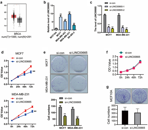
Figure 2. Knockdown of LINC00665 inhibited the migration and invasion and induced the apoptosis in breast cancer cells.
Transwell assay was performed to detect the migration and invasion in MCF7 (A) and MDA-MB-231 (B) cells. (C, D) Flow cytometry was performed to analysis the apoptosis of si-LINC00665 and si-con cells. (E) The expression of apoptosis-related proteins Bcl-2, Bax, and Cleaved caspase 3 was detected using Western blot. **P < 0.01.
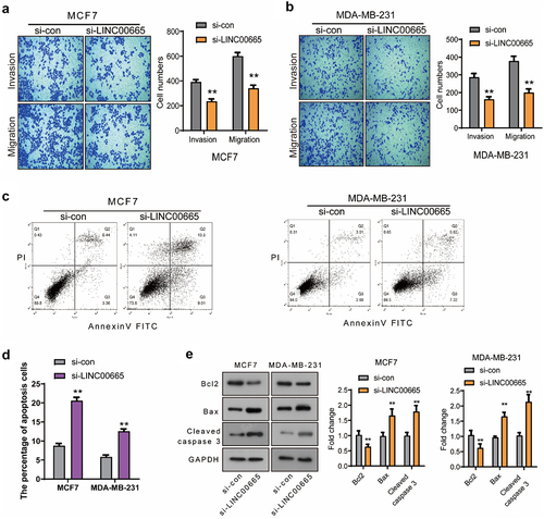
Figure 3. Knockdown of LINC00665 inactivated the AKT/mTOR signaling pathway in breast cancer cells.
(A) The expressions of p-AKT, AKT, p-mTOR, and mTOR were analyzed using Western blot. The levels of p-AKT/AKT and p-mTOR/mTOR in MCF7 (B) and MDA-MB-231 (C) cells were analyzed using ImageJ. **P < 0.01.
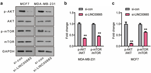
Figure 4. LINC00665 promoted the expression of SRCAP through sponging miR-641 in breast cancer cells.
(A) MiRNAs, which could bind to LINC00665, were analyzed using the online software StarBase. The sequence of LINC00665 was constructed into pGL3 plasmid (LINC00665-wt), and then dual-luciferase report gene analysis was performed to detect the combination of LINC00665 and miRNAs (miR-542-3p, miR-624-5p, miR-641, miR-425-5p, and miR-30-3p). (B) The sequence of LINC00665 with mutation-binding site (UACAGAA) was constructed into pGL3 plasmid to generate LINC00665-mut plasmid (upper panel). The combination of LINC00665 and miR-641 was further confirmed using dual-luciferase report gene analysis (bottom panel). (C) The miR-641 level was detected using qPCR in si-LINC00665 and si-con group. (D) Potential targets of miR-641 were analyzed using the online software StarBase. qPCR was performed to detect the levels of miR-641ʹs potential targets (SRCAP, SIKE1, NADK, KHDC4, and HSPG2) after the transfection of miR-641 mimics. (E) The sequence of SRCAP-3ʹUTR with wild-type (UGUCUU) or mutation-binding site (ACAGAA) was constructed into pGL3 plasmid to generate SRCAP-wt or SRCAP-mut plasmid, respectively (upper panel). The combination of SRCAP and miR-641 was confirmed using dual-luciferase report gene analysis (bottom panel). (F) The SRCAP level was detected using qPCR after the transfection of miR-641 mimics with the untreated MCF7 and MDA-MB-231 cells as control groups. *P < 0.05; **P < 0.01.
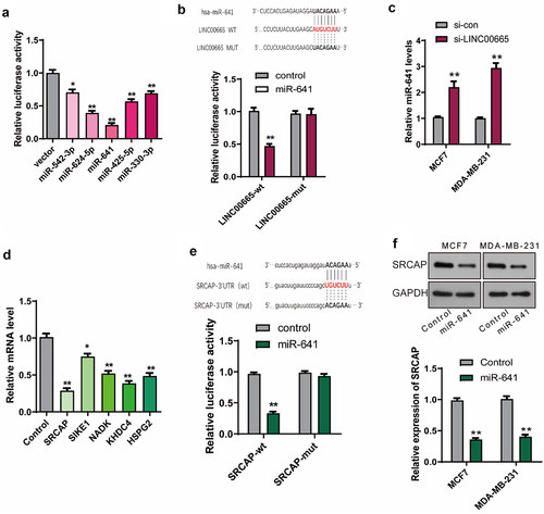
Figure 5. Knockdown of LINC00665 inhibited the proliferation and invasion through down-regulating SRCAP by sponging miR-641.
(A) CCK8 was performed to detect the proliferation in MCF7 and MDA-MB-231 cells. (B and C) Transwell assay was used to detect the invasion in MCF7 and MDA-MB-231 cells.
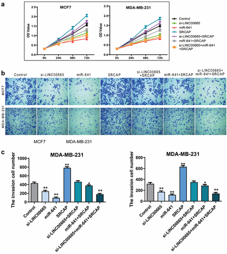
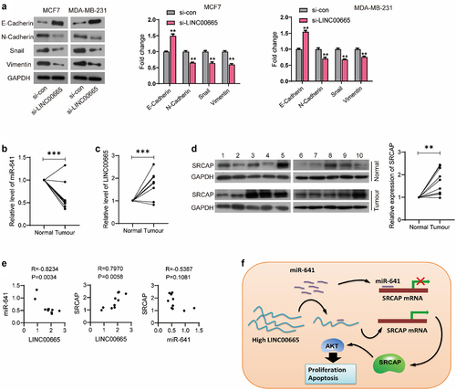
Figure 6. LINC00665 was up-regulated in breast cancer patients.
(A) The expression of EMT makers were detected using Western blot. (B) The expression of LINC00665 in clinical samples of 10 breast cancer patients was detected using qPCR. (C) The level of miR-641 was also detected using qPCR. (D) SRCAP expression was determined using Western blot. (E) The correlation between the expression of LINC00665, miR-641, and SRCAP in tumor tissues was evaluated using Pearson Correlation Coefficient. (F) Schematic diagram of LINC00665/miR-641/SRCAP axis promoting tumor cell proliferation. **P < 0.01; ***P < 0.001.