
Figures & data
Figure 1. MiR-885-3p is low-expressed in asthma patients’ plasma and 16HBE cells stimulated by LPS.
(a) The heat map shows the differential expression of miRNAs in normal and asthmatic bronchial epithelial cells.(b,c) Detection of miR-885-3p expression in asthma patients’ plasma and 16HBE cells stimulated by LPS via qRT-PCR.(d) ELISA was performed to detect the content of TNF-α, IL-8, and IL-6 in LPS-stimulated 16HBE cell supernatant.All of the experiments were performed in triplicate. Student’s t test, *P < 0.05, and ***P < 0.001.
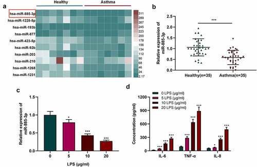
Figure 2. MiR-885-3p alleviates LPS-stimulated 16HBE cell inflammation.
(a) Detection of miR-885-3p expression in 16HBE cells transfected with miR-NC or miR-885-3p mimics via qRT-PCR.(b) qRT-PCR was performed to detect the relative miR-885-3p expression in 16HBE cells after transfection of miR-885-3p mimic or stimulated with 10 μg/ml LPS.(c–e) ELISA was performed to measure IL-6, TNF-α and IL-8 concentrations in the supernatant of 16HBE cells stimulated with 10 μg/ml LPS or transfected with miR-885-3p mimic.All of the experiments were performed in triplicate. Student’s t test, **P < 0.01 and ***P < 0.001.
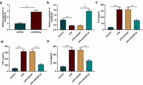
Figure 3. The impacts of miR-885-3p on 16HBE cell viability and apoptosis.
(a–c) CCK-8 assay and flow cytometry were conducted to evaluate the viability (A) and apoptosis rate (B, C) of 16HBE cells transfected with miR-885-3p mimic or treated with 10 μg/ml LPS, respectively.(d,e) Detection of the cleaved caspase-3, Bax and Bcl-2 levels in 16HBE cells, which were stimulated with 10 μg/ml LPS or transfected with miR-885-3p mimic, via Western blotting.All of the experiments were performed in triplicate. Student’s t test, **P < 0.01, and ***P < 0.001.
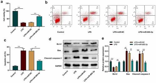
Figure 4. TLR4 is miR-885-3p’s downstream target.
(a) The binding sequence of TLR4 mRNA 3’-UTR with miR-885-3p.(b) The targeted relationship of miR-885-3p with TLR4 was verified through dual-luciferase assay.(c,d) Western blot and qRT-PCR were utilized for detecting TLR4 mRNA and protein expression in 16HBE cells with transfection of miR-885-3p mimic.(e,f) Western blotting and qRT-PCR were conducted to detect TLR4 mRNA and protein expression in 16HBE cells stimulated with LPS at different concentrations (5, 10, and 20 μg/ml).All of the experiments were performed in triplicate. Student’s t test, *P < 0.05, **P < 0.01, and ***P < 0.001.
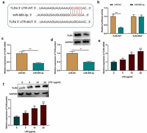
Figure 5. MiR-885-3p suppresses LPS-induced cell inflammatory injury through targeting TLR4.
(a,b) Western blotting and qRT-PCR were conducted to examine TLR4 mRNA and protein expression in 16HBE cells with transfection of miR-885-3p mimic or stimulated with 10 μg/ml LPS or co-transfected with TLR4 overexpression plasmid and miR-885-3p mimic.(c–e) ELISA was performed to detect TNF-α, IL-8 and IL-6 levels in 16HBE cell culture supernatant.(f,g) 16HBE cell viability (F) and apoptosis rate (G) were evaluated via CCK-8 and flow cytometry.(h,i) Cleaved caspase-3, Bax and Bcl-2 expression levels in 16HBE cells were detected by Western blotting.All of the experiments were performed in triplicate. Student’s t test, *P < 0.05, **P < 0.01, and ***P < 0.001.
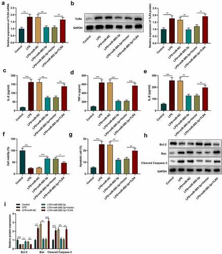
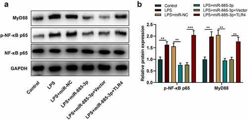
Figure 6. MiR-885-3p suppresses the NF-κB-MyD88 signaling activation.
(a,b) Western blotting was utilized for detecting p-NF-κB p65, MyD88 and NF-κB p65 expression in 16HBE cells with transfection of miR-885-3p mimics or treated with 10 μg/ml LPS or co-transfected with TLR4 overexpression plasmid and miR-885-3p mimic.All of the experiments were performed in triplicate. Student’s t test, *P < 0.05, and **P < 0.01.
Supplemental material
Supplemental Material
Download MS Word (2.8 MB)Data availability statement
The data for supporting the findings of the present study are available upon request from the corresponding authors.