Figures & data
Figure 1. PB2 inhibited viability and promoted apoptosis of OSCC cells. (a) The chemical structure of PB2. (b) The viability of oral mucosa epithelial cell (OMEC) treated with PB2 was detected by CCK-8 assay. (c) The viability of human OSCC cell line (SCC-25) treated with PB2 was detected by CCK-8 assay. (d) The apoptosis of SCC-25 cells treated with PB2 was analyzed by Tunel assay. (e) The expression of apoptosis related proteins in SCC-25 cells treated with PB2 was determined by Western blot. *P < 0.05, **P < 0.01 and ***P < 0.001 vs. PB2 (0 μM) group.
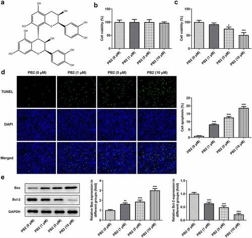
Figure 2. PB2 inhibited invasion, migration and epithelial-mesenchymal transition (EMT) of OSCC cells. The migration (a) and invasion (b) of SCC-25 cells treated with PB2 were detected by wound healing assay and transwell assay. The expression of metastasis associated proteins (c) and EMT related proteins (d) was analyzed by Western blot. *P < 0.05, **P < 0.01 and ***P < 0.001 vs. PB2 (0 μM) group.
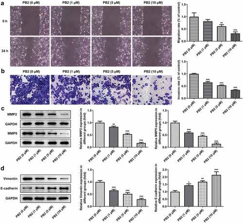
Figure 3. PB2 inhibited VEGF/VEGFR2 signaling and tumor angiogenesis in OSCC. (a) The related genes of PB2 were analyzed by STTICH database. (b/c) The expression of VEGF/VEGFR2 signaling in SCC-25 cells treated with PB2 was analyzed by Western blot. (d) The angiogenesis of SCC-25 cells treated with PB2 was detected by tube formation assay. *P < 0.05 and ***P < 0.001 vs. PB2 (0 μM) group.
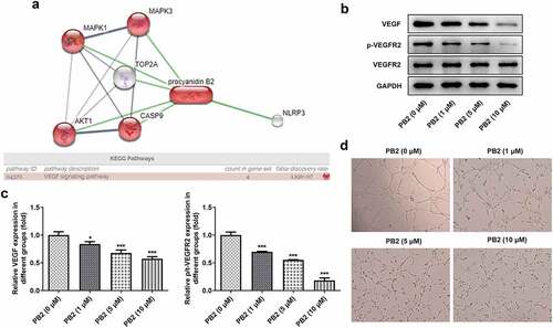
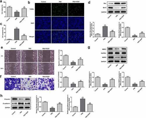
Figure 4. Activation of VEGF/VEGFR2 signaling reduced the effect of PB2 on growth and metastasis of OSCC cells. (a) The viability of SCC-25 cells treated with PB2 with or without VEGF was detected by CCK-8 assay. The apoptosis (B/C) and related proteins (d) in SCC-25 cells treated with PB2 with or without VEGF were respectively analyzed by Tunel assay and Western blot. The migration (e) and invasion (f) of SCC-25 cells treated with PB2 with or without VEGF were detected by wound healing assay and transwell assay. The expression of metastasis associated proteins (g) and EMT related proteins (h) was analyzed by Western.